. 2025 Apr 29;16(1):94.
doi: 10.1186/s13244-025-01945-3.

Radiological approach to metatarsalgia in current practice: an educational review

Affiliations

Radiological approach to metatarsalgia in current practice: an educational review

Océane Palka et al. Insights Imaging. .

Abstract

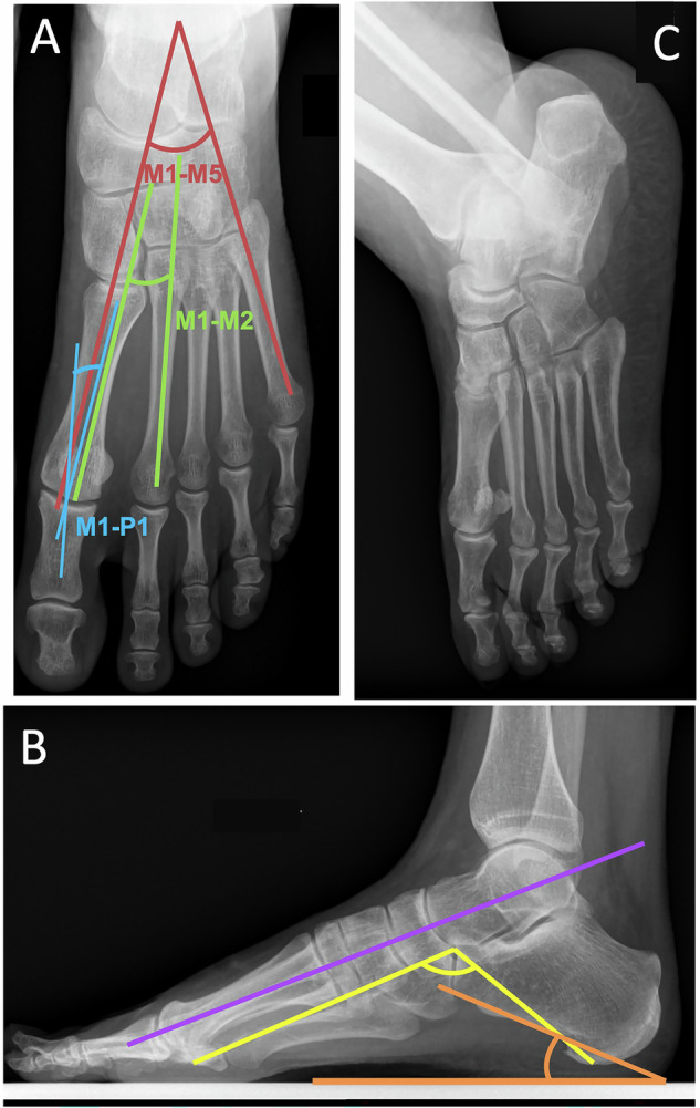
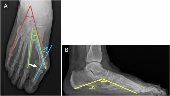
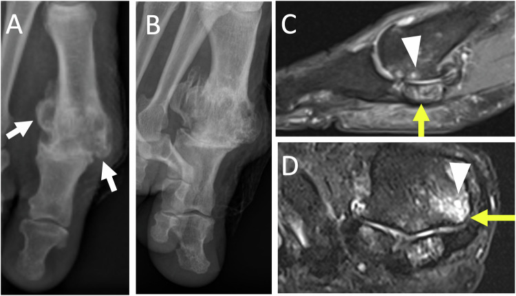
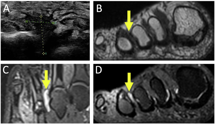
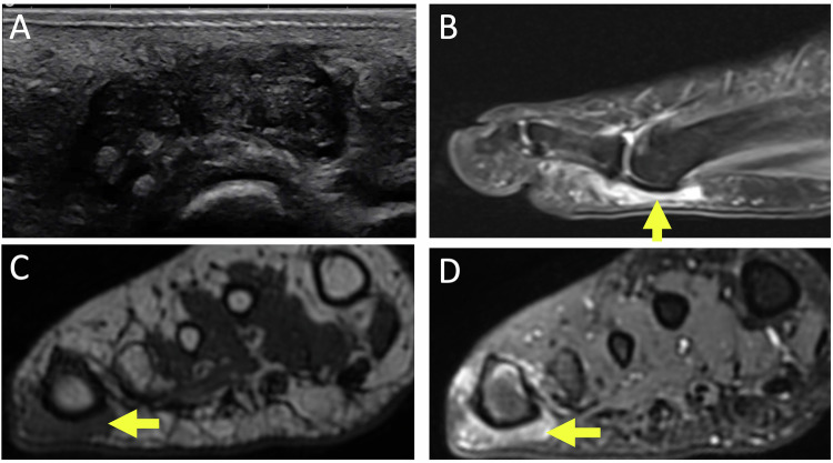
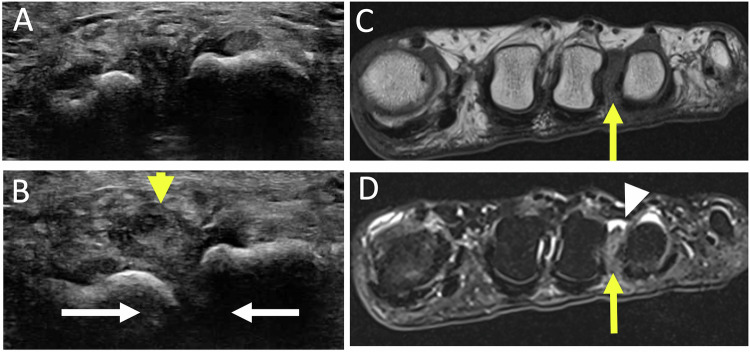
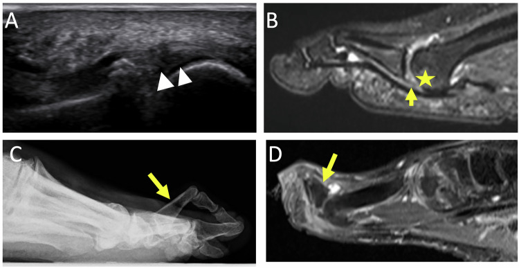
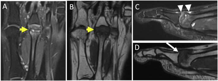
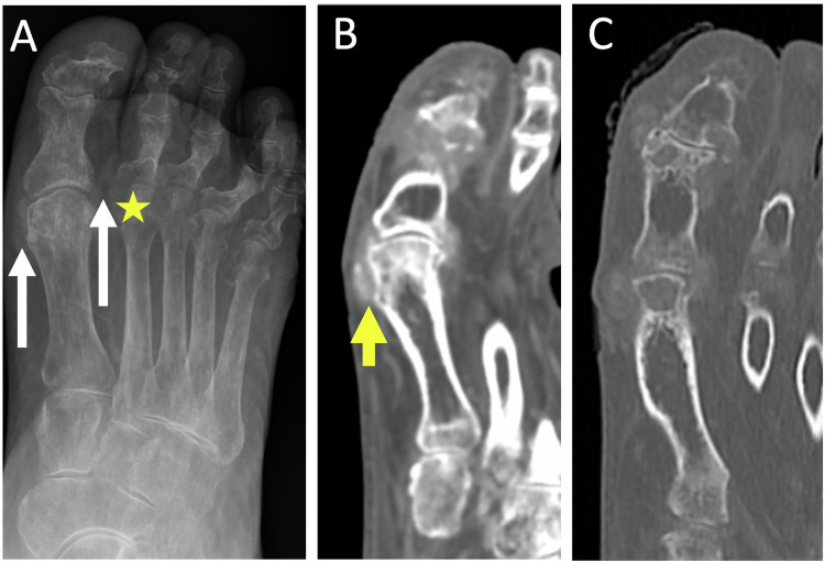
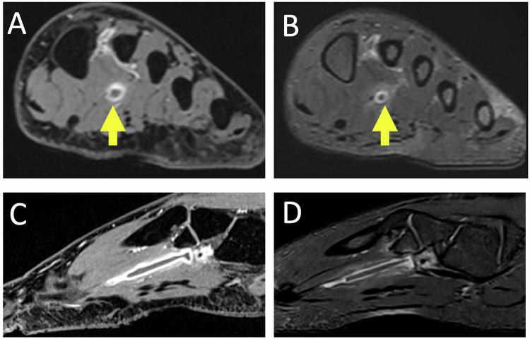
Metatarsalgia, characterized by forefoot pain, is frequent and is primarily due to foot static disorders. Initial evaluation with weight-bearing radiographs is essential, allowing precise analysis of the architecture of the foot. Ultrasound is useful for soft tissue and tendon examination and provides the best clinical correlation. Computed Tomography provides detailed bone assessment and is helpful for pre-operative planning. Magnetic Resonance Imaging is the gold standard modality, offering superior soft tissue contrast. The common causes of metatarsalgia include hallux pathologies (hallux valgus, hallux rigidus, and sesamoid issues), bursitis (intermetatarsal and subcapitellar), Morton's neuroma, second ray syndrome, stress fractures, and systemic pathologies affecting the foot. Combining clinical and imaging data is crucial for accurate diagnosis and effective management of metatarsalgia. Post-traumatic causes of metatarsalgia are beyond the scope of this article and will not be described. CRITICAL RELEVANCE STATEMENT: Metatarsalgia, the pain of the forefoot, necessitates accurate imaging for diagnosis and management. This review critically assesses imaging techniques and diagnostic approaches, aiming to enhance radiological practice and support effective therapeutic decision-making. KEY POINTS: Metatarsalgia commonly results from foot static disorders, requiring weight-bearing radiographs for assessment. MRI is often the gold standard examination, but ultrasound is complementary, allowing for a radioclinical approach with dynamic examinations. The radiologist is crucial in diagnosing metatarsalgia, providing essential imaging, and guiding treatment.

Keywords: Forefoot; MRI; Metatarsalgia; Radiograph; Ultrasound.


Conflict of interest statement

Declarations. Ethics approval and consent to participate: Not applicable. Consent for publication: University Hospitals of Angers and Rennes guidelines do not require individual consent for the publication of images, as long as they are anonymized. Competing interests: The authors declare that they have no known competing interests.

Figures

Fig. 1
Fig. 1
Main radiological views of the forefoot with measurements used in current practice. Weight-bearing dorso-plantar (A) and lateral (B) views and medial oblique view (C). In dorsoplantar view (A)—the metatarsophalangeal angle of the hallux: N < 15° (blue angle)—the metatarsus varus angle: N < 10° (around 5°) (green angle)—the angle of opening of the forefoot (width of the metatarsal fan): N between 15° and 20° (red angle). In profile view (B)—the Djian–Annonier’s angle (angle of the medial arch of the foot): N between 120° and 130° (yellow angle)—the talo metatarsal angle (Meary–Tomeno angle): normally forming a straight line (purple line). The slope of the calcaneus: N between 15° and 25° (orange angle)
Fig. 2
Fig. 2
Hallux valgus of the right foot in a 63-year-old woman with a flat foot complicated by metatarsophalangeal osteoarthritis. Weight-bearing dorsoplantar (A) and lateral (B) radiographic views of the right foot. Forefoot opening angle (red angle): 32°; metatarsus varus angle (green angle): 18°; metatarsophalangeal angle of the first ray (blue angle): 34°; Toe formula: square foot; Djian–Annonier angle (yellow angle): 135° Degenerative arthropathy of the MTPJ (arrow), with moderate narrowing of the joint space, some subchondral bone changes of the condensing and microgeodic type, and a small amount of marginal osteophyte production
Fig. 3
Fig. 3
Chronic plantar pain of the first ray in the woman of 23 years old. Radiographs of the forefoot in weight-bearing dorso plantar (A) and sesamoid (B) views show a fragmented appearance of lateral sesamoid suggesting osteonecrosis (long white arrows). Unexplained right foot pain for several months in a 65-year-old woman. Sagittal MR images show increased signal intensity in T2-weighted (C) and decreased signal intensity in T1-weighted (D) of the posterior part of the bipartite medial sesamoid (short yellow arrows), associated with reactive synovitis and soft tissue inflammation (arrowhead), in favor of medial hallux sesamoiditis
Fig. 4
Fig. 4
Hallux rigidus in a 66-year-old man. Radiographics in weight-bearing dorso plantar view (A) and medial oblique view (B) show severe metatarsophalangeal osteoarthritis of the first ray, with pinching of the joint, marginal osteophytosis (white arrows). Sagittal (C) and axial (D) MR images in T2-weighted fat-suppressed show joint effusion, microgeodic subchondral lesion (arrowheads) on the medial side of the base of the proximal phalanx and on the medial side of the head of the first metatarsal with associated metatarso sesamoid osteoarthritis with significant subchondral edema (yellow arrows)
Fig. 5
Fig. 5
Intermetatarsal bursitis of the third space in a 48-year-old man presenting with right fourth toe pain aggravated by prolonged standing. US (A) shows a hypoechogenic mass of the third space which compresses in. MR images show a hypointense intermetatarsal mass in the T1-weighted short axis (B) and fluid hyperintense mass in the T2-weighted long and short axis (C, D) (arrows)
Fig. 6
Fig. 6
Submetatarsal bursitis in a 73-year-old woman with chronic left metatarsalgia. US (A) shows a hypoechoic edematous infiltration of the subcapito-metatarsal area of the fourth ray. MR images show hypointense in the T1-weighted short axis (C) and fluid hyperintense mass in the T2-weighted long and short axis (BD) (arrows)
Fig. 7
Fig. 7
Morton’s neuroma in a 76-year-old woman presenting chronic left metatarsalgia with paresthesia of the fourth ray. US in short axis (A, B) shows hypoechoic mass located in the plantar intermetatarsal space, which dislocate (short arrow) during dynamic lateral compression (long white arrows) of the forefoot: Mulder’s sonographic sign MR images of the forefoot in short axis show oval mass in the third intermetatarsal space (long arrows), hypointense in T1-weighted image (C), hyperintense in T2-weighted image (D), overlain by a bursitis (arrowhead)
Fig. 8
Fig. 8
Second ray syndrome in a 34-year-old woman presenting with pain between the first and second intermetatarsal spaces for two months. US in sagittal plantar view (A) shows plantar plate irregularity (arrowheads), synovitis, and effusion of the 2nd MTP. Sagittal fat-suppressed T2-weighted MR image (B) shows rupture of the plantar plate (short arrow), with dorsal subluxation of P1, metatarsophalangeal synovitis with subchondral bone hypersignal of the head of the second metatarsal on its plantar side (star). Second ray syndrome in a 70-year-old man in terminal phase. Radiograph in lateral view (C) and sagittal MR image in T2-weighted (D) show dorsal dislocation of P1 (long arrows)
Fig. 9
Fig. 9
Stress fracture of the right second metatarsal in a 17-year-old patient. Radiograph in dorsoplantar view (A) taken retrospectively one month later, shows periosteal appositions of the second right metatarsal (arrowheads). A stress fracture in a 68-year-old man. MRI images show edematous intraosseous signal abnormalities in the proximal third of the shaft of the 4th metatarsal, surrounding a serpiginous hypo signal linear image (arrow) in T1 (B) and T2 (C): the appearance of a semi-recent stress fracture
Fig. 10
Fig. 10
Freiberg’s disease of the second metatarsal of the left foot in the late degenerative stage in a 21-years-old woman. MR images in T2-weighted fat-suppressed (AC) and T1-weighted (BD) show deformity and hypertrophy of the head of the second metatarsal, edematous bone signal abnormalities (yellow short arrows) with geodic subchondral bone remodeling of the head, marginal osteophytosis of the base of proximal phalanx (long arrow), and intra-articular metatarsophalangeal effusion (arrowheads)
Fig. 11
Fig. 11
Chronic tophaceous gout in an 86-year-old man. Radiograph in dorso-plantar view (A) shows gouty tophus of the periarticular soft tissues opposite the MTPJ of the first and second rays (long arrows), bone erosion of the medial edge of the head of the second metatarsal (star), distal interphalangeal arthropathy. The MTPJ line spacing of the first ray is relatively intact compared to the extent of the erosions. CT (B, C) shows soft tissue thickening over the MTPJ of the hallux, containing a few discrete calcifications suggestive of gouty tophus (short arrow)
Fig. 12
Fig. 12
Plantar medial venous thrombosis in a 52-year-old woman presenting with pain in the foot for five days. MR images of the forefoot in T1 weighted fat-suppressed post gadolinium short and sagittal axis (AC) and T2-weighted fat-suppressed (BD) show acute thrombosis with venous defect in the medial plantar vein (arrows) with perivascular edema

Similar articles

References

    1. Halstead J, Munteanu SE (2023) Current and future advances in practice: mechanical foot pain. Rheumatol Adv Pract 7:rkad081 - PMC - PubMed
    1. Guerini H, Morvan G, Mathieu P et al (2014) Métatarsalgies et imagerie: algorithme diagnostique. Rev Rhum Monogr 81:138–146
    1. Thomas JL, Kunkel MW, Lopez R, Sparks D (2006) Radiographic values of the adult foot in a standardized population. J Foot Ankle Surg 45:3–12 - PubMed
    1. Joong MA, El-Khoury GY (2007) Radiologic evaluation of chronic foot pain. Am Fam Phys 76:975–983 - PubMed
    1. Djian A, Annonier C, Denis A, Baudoin P(1968) Radiopodometry (principles and results)] J Radiol Electrol Med Nucl 49:769–772 - PubMed

LinkOut - more resources