. 2025 Apr 29;16(1):3974.
doi: 10.1038/s41467-025-57843-1.

Mechanistic studies of mycobacterial glycolipid biosynthesis by the mannosyltransferase PimE

Affiliations

Mechanistic studies of mycobacterial glycolipid biosynthesis by the mannosyltransferase PimE

Yaqi Liu et al. Nat Commun. .

Abstract

Tuberculosis (TB), a leading cause of death among infectious diseases globally, is caused by Mycobacterium tuberculosis (Mtb). The pathogenicity of Mtb is largely attributed to its complex cell envelope, which includes a class of glycolipids called phosphatidyl-myo-inositol mannosides (PIMs). These glycolipids maintain the integrity of the cell envelope, regulate permeability, and mediate host-pathogen interactions. PIMs comprise a phosphatidyl-myo-inositol core decorated with one to six mannose residues and up to four acyl chains. The mannosyltransferase PimE catalyzes the transfer of the fifth PIM mannose residue from a polyprenyl phosphate-mannose (PPM) donor. This step contributes to the proper assembly and function of the mycobacterial cell envelope; however, the structural basis for substrate recognition and the catalytic mechanism of PimE remain poorly understood. Here, we present the cryo-electron microscopy (cryo-EM) structures of PimE from Mycobacterium abscessus in its apo and product-bound form. The structures reveal a distinctive binding cavity that accommodates both donor and acceptor substrates/products. Key residues involved in substrate coordination and catalysis were identified and validated via in vitro assays and in vivo complementation, while molecular dynamics simulations delineated access pathways and binding dynamics. Our integrated approach provides comprehensive insights into PimE function and informs potential strategies for anti-TB therapeutics.


Conflict of interest statement

Competing interests: The authors declare no competing interests.

Figures

Fig. 1
Fig. 1. Biosynthetic pathway and structural architecture of PimE.
a Overview of PIM biosynthesis in mycobacteria, featuring the enzymatic reactions carried out by key mannosyltransferases. Enzyme names are indicated above the reaction arrows. The early steps occur on the cytoplasmic side of the plasma membrane, where PimA and PimB transfer mannose residues from GDP-mannose to the 2-OH and 6-OH positions of the inositol moiety of phosphatidylinositol (PI), yielding PIM1 and PIM2, respectively. PatA then acylates the 6-OH position of the mannose ring of PIM2, resulting in AcPIM2. An unknown acyltransferase can add an additional acyl chain to the 3-OH of the inositol of AcPIM2 to form Ac2PIM2. These acylated mannosylated intermediates are presumed to translocate to the outer leaflet of the membrane by an unknown flippase. PimC and PimD are thought to catalyze the subsequent tri- and tetra-mannosylation steps, leading to the formation of Ac1PIM4. PimE catalyzes the transfer of the fifth mannose residue from polyprenyl-monophospho-β-d-mannose (PPM) to Ac1PIM4, forming Ac1PIM5. The enzyme responsible for the conversion of Ac1PIM5 to AcPIM6 remains unknown. b Mannosyl transfer reaction catalyzed by PimE, showing the formation of an α(1 → 2) glycosidic bond between PPM and Ac1PIM4, yielding Ac1PIM5 and PP. Only tri-acylated PIMs (Ac1PIM4 and Ac1PIM5) are shown for the sake of simplicity. c Cryo-EM map of MaPimE in complex with Fab-E6. PimE is depicted in rainbow, while the Fab-E6 is shown in gray. (d) PimE is shown as a ribbon colored in rainbow, as in Fig. 1f. e TM helix arrangement of PimE is depicted as a cross-section colored in a rainbow as in Fig. 1f. f Topological diagram of PimE showing the arrangement of TM segments and extracellular domains. The key catalytic residue D58 is shown as a red dot.
Fig. 2
Fig. 2. The putative substrate-binding cavity of PimE.
a The putative active site cavity within PimE is colored in semitransparent yellow, showing its elongated, cashew-like shape and orientation relative to the membrane plane. b, c PimE viewed parallel (b) and perpendicular (c) to the membrane plane, with the elongated, cashew-shaped cavity shown in yellow. The cavity is oriented almost parallel to the membrane plane, with its two ends (Cavity End 1, CE1, and Cavity End 2, CE2) curving slightly toward the membrane. The cavity is surrounded by periplasmic loops (PL1 to PL3) and the connecting loops between TM helices 3 and 4 and TM helices 9 and 10. df PimE rendered in surface representation colored by hydrophobicity on a purple (very hydrophilic) to tan (very hydrophobic) scale (d) by conservation on a cyan (low conservation) to magenta (absolute conservation) scale (e) and by electrostatic potential (f) on a range of ±5 kBT/e. The putative reaction center at the central part of the cavity is marked with a dotted circle.
Fig. 3
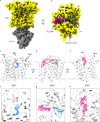
Fig. 3. The cryo-EM structure of substrate-bound MaPimE.
a, b Cryo-EM density for the product-bound complex of MaPimE with Ac1PIM5 (product) and PP (by-product), viewed parallel to the membrane plane. Fab-E6 binds to the same cytoplasmic domain of MaPimE as observed in the apo structure. ce Ribbon representation for the product-bound structure of PimE (white) bound with PP (blue) and Ac1PIM5 (magenta) shown in different orientations. PP and Ac1PIM5 adopt a curved shape, with their polar head groups projecting into the hydrophilic center of the substrate-binding cavity. fh A focused view of the region where PP and Ac1PIM5 bind. PP and Ac1PIM5 adopt a curved shape. The polyprenyl chain of PP points towards the hydrophobic TM domain groove composed of TM helices 6 and 9, while Ac1PIM5 is surrounded by PL1, JM1, and JM4, with its acyl chains likely extending toward the TM region near TM helix 3.
Fig. 4
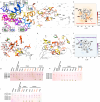
Fig. 4. Structural insights into MaPimE: Active site, substrate interactions, and functional residues.
a Close-up view of the active site from the cryo-EM product-bound structure of PimE bound with products Ac1PIM5 and by-product PP. Key residues are shown as sticks. Insets: Sequence logos highlighting the conservation of active site residues involved in interactions with PP or Ac1PIM5. b Same view as (a) with residues colored according to mutational effects: red-orange for complete activity loss, yellow for reduced activity, and green for no change in activity. c, d RoseTTAFold docked models of PimE with full-length donor PPM and acceptor Ac1PIM4. Residues are colored as in (b) based on mutational effects. Hydrogen bonds are shown as black dotted lines in panels (ad). e, f Density of PPM (e) and Ac1PIM4 (f) in CG-MD simulations with respect to PimE, with the protein backbone shown as gray lines. Regions of darker color show higher density by the lipid. Key areas of the protein are highlighted. gi HPTLC analysis of PIMs profiles from M. smegmatis strains. Each plate compares PIMs from WT M. smegmatis mc²155, ΔpimE mutant strain, and ΔpimE complemented with WT or mutant MaPimE. Major PIM species are indicated. Yellow stars mark mutants with reduced but not abolished activity (detectable PIM6 production). Mutations causing complete loss of activity are labeled in red-orange. All experiments were independently repeated two times with similar results.
Fig. 5
Fig. 5. Proposed catalytic mechanism of PimE.
a Schematic representation of the active site of PimE with bound substrates, showing the positioning of PPM and Ac1PIM4. D58, located at the tip of JM1, acts as the catalytic base. b Proposed catalytic mechanism, showing the role of D58 in deprotonating the 2-OH group of Ac1PIM4, initiating a nucleophilic attack on the anomeric carbon of PPM. This leads to the formation of an α(1 → 2) glycosidic bond between the mannose moiety of PPM and Ac1PIM4. The phosphate group of PPM is cleaved and stabilized by K195 and H322, forming the product Ac1PIM5 and the by-product PP. The tetra-acylated acceptor Ac2PIM4 and its corresponding glycosylated product Ac2PIM5 are not shown here for the sake of simplicity.

Update of

References

    1. Daniel, T. M. The history of tuberculosis. Respir. Med.100, 1862–1870 (2006). - PubMed
    1. Wirth, T. et al. Origin, spread and demography of the Mycobacterium tuberculosis complex. PLoS Pathog.4, e1000160 (2008). - PMC - PubMed
    1. World Health Organization. 2024 Global Tuberculosis Report (WHO, 2024).
    1. Cirillo, J. D. & Kong, Y. Tuberculosis Host-Pathogen Interactions (Springer International Publishing, 2019).
    1. McNeil, M. R. & Brennan, P. J. Structure, function and biogenesis of the cell envelope of mycobacteria in relation to bacterial physiology, pathogenesis and drug resistance; some thoughts and possibilities arising from recent structural information. Res. Microbiol.142, 451–463 (1991). - PubMed

MeSH terms

LinkOut - more resources