. 2025 Apr 29;16(1):4028.
doi: 10.1038/s41467-025-59377-y.

Engineering of extracellular vesicles for efficient intracellular delivery of multimodal therapeutics including genome editors

Affiliations

Engineering of extracellular vesicles for efficient intracellular delivery of multimodal therapeutics including genome editors

Xiuming Liang et al. Nat Commun. .

Abstract

Intracellular delivery of protein and RNA therapeutics represents a major challenge. Here, we develop highly potent engineered extracellular vesicles (EVs) by incorporating bio-inspired attributes required for effective delivery. These comprise an engineered mini-intein protein with self-cleavage activity for active cargo loading and release, and fusogenic VSV-G protein for endosomal escape. Combining these components allows high efficiency recombination and genome editing in vitro following EV-mediated delivery of Cre recombinase and Cas9/sgRNA RNP cargoes, respectively. In vivo, infusion of a single dose Cre loaded EVs into the lateral ventricle in brain of Cre-LoxP R26-LSL-tdTomato reporter mice results in greater than 40% and 30% recombined cells in hippocampus and cortex respectively. In addition, we demonstrate therapeutic potential of this platform by showing inhibition of LPS-induced systemic inflammation via delivery of a super-repressor of NF-ĸB activity. Our data establish these engineered EVs as a platform for effective delivery of multimodal therapeutic cargoes, including for efficient genome editing.


Conflict of interest statement

Competing interests: O.W., J.Z. N., D.G. A.G., M.J.A.W. and S.E.-A. are consultants and stakeholders in Evox Therapeutics Limited, Oxford, United Kingdom. A.D.F., and J.H. are employees of Evox Therapeutics Limited, Oxford, United Kingdom. Evox Therapeutics filed a patent application related to the data used in this work. The patent was applied by Evox Therapeutics Limited. The inventors are Xiuming Liang, Dhanu Gupta, Samir EL Andaloussi and Joel Nordin. Its application number is PCT/EP22/84310 which is still under evaluation. The development of VEDIC and VFIC systems of this work was included in this patent. All the data are available in the manuscript or in the supplemental information. Materials are available upon signing the material transfer agreement (MTA) submitted to S.E.-A. and Evox Therapeutics Limited, Oxford, United Kingdom. The remaining authors declare no competing interests.

Figures
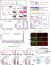
Fig. 1
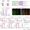
Fig. 1. Development of the VEDIC system for high efficiency intracellular protein delivery by EVs.
a Graphic abstract of the development of VEDIC and VFIC systems for high efficiency intracellular protein delivery in vitro and in vivo. Intein in tripartite fusion protein (EV-sorting Domain-Intein-Cargo) performs C-terminal cleavage during the process of EV-biogenesis, resulting in free cargo protein inside EVs. Together with a fusogenic protein, e.g. VSV-G, these engineered EVs achieve efficeint intracellular delivery of cargo protein (Cre and super repressor of NF-ĸB) or protein/RNA complex (Cas9/sgRNA RNPs) both in reporter cells and in mice models. b Fluorescence reporter construct expressed in the reporter cells generated to measure Cre delivery. c Constructs used for developing the VEDIC system. d Schematic of intein cleavage and intraluminal cargo release during EV biogenesis, MVB: multivesicular body. e Percentage of GFP positive reporter cells after incubating EVs for 2 days, as evaluated by flow cytometry. f Screen of fusogenic proteins co-expressed with CD63-Intein-Cre for EV-mediated delivery analyzed in T47D-TL cells after a two-day incubation period. g, Percentage of GFP positive reporter cells after exposure to EVs derived from VSV-G co-transfected cells. h EV dose-and time-dependent recombination in HeLa-TL reporter cells mediated by VEDIC EVs. i, EV dose-and time-dependent recombination in B16F10-TL reporter cells mediated by VEDIC EVs. j Cre and VSV-G protein was analyzed in T47D-TL reporter cells by Western blot (WB) analysis, 48 hours (h) after addition of engineered EVs loaded with Cre in 24-well plates. k Comparison of the Cre transfer efficiency between VEDIC EVs and published Nanoblade system. Two-way ANOVA (Tukey) multiple comparisons test was used for analysis of (g) (i) and (k).; One-way ANOVA (Tukey) multiple comparisons test was used for analysis of (f). Experiments were done with 3 biological replicates except the Nanoblade (Cre) particles in (k) which had 2 biological replicates. Data are shown as mean ± SD. a, d and right panel of k Created in BioRender.com, with attribution line Zheng, W. (2025) https://BioRender.com/n11l657, Zheng, W. (2025) https://BioRender.com/i50r712 and Zheng, W. (2025) https://BioRender.com/n32h319 respectively. Exact statistical analysis was reported in the Source data and Source data are provided as a Source Data file.
Fig. 2
Fig. 2. Development of the VFIC system to further improve intracellular protein delivery by EVs.
a VSV-G+ and CD63 + EV concentrations as determined by single-vesicle flow cytometry after transfection with VSV-G-mNG alone or VSV-G-mNG and CD63 together. b Constructs generated for developing the VFIC system. The last construct was generated as a negative control where Cre was replaced with the bacteriophage protein MS2. c EV dose-dependent recombination in B16F10-TL cells mediated by VSV-G-Foldon-Intein-Cre and VSV-G-Intein-Cre EVs as evaluated by flow cytometry. d Representative images showing GFP positive HeLa-TL cells 24, 48 and 72 h after exposure to VFIC EVs at different doses. Scale bar, 100 µm. eh Recombination in hard-to-transfect reporter cells (MSC-TL, THP-1-TL, Raw264.7-TL and K562-TL) mediated by VFIC EVs after 48 h. i The efficiency of Cre transfer in reporter cells by EVs isolated using TFF + UF + SEC method. TFF: tangential flow filtration; UF: ultrafiltration; SEC: size exclusion chromatography. j Dynamic Cre delivery in HeLa-TL cells using VFIC EVs purified by DGUC. DGUC: density gradient ultracentrifugation; T1-T3: technical replicates. Two-way ANOVA (Tukey) multiple comparisons test was used for analysis of (c), (eh) and (i). Experiments were done with 3 biological replicates and data are shown as mean ± SD. Exact statistical analysis was reported in the Source data and Source data are provided as a Source Data file.
Fig. 3
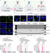
Fig. 3. The pH-sensitive intein performs C-terminal cleavage during EV-biogenesis.
a Schematic illustration of the cleavage mechanism of different engineered intein variants. b Protein expression of different engineered mutant intein constructs in whole cell lysates (WCL) and isolated EVs derived from HEK293T cells evaluated by western blot analysis. Lysates from 5 × 105 EV-producing cells and 1×10¹⁰ engineered EVs were loaded onto the western blot gel. TSG101, syntenin-1 and β-actin were used as EV markers and Calnexin was used as a cellular organelle marker (endoplasmic reticulum) that should be absent in EV samples. ce Recombination in reporter cells mediated by EVs derived from engineered cells using mutant inteins (H439Q and N440A) in VEDIC system. Two-way ANOVA (Tukey) multiple comparisons test was used for analysis of (ce). Experiments were done with 3 biological replicates and data are shown as mean ± SD. a Created in BioRender.com, Zheng, W. (2025) https://BioRender.com/v41e325. Exact statistical analysis was reported in the Source data and Source data are provided as a Source Data file.
Fig. 4
Fig. 4. VSV-G boosts endosomal escape following receptor-mediated endocytosis of engineered EVs into recipient cells.
a, b Properties of the two VSV-G mutants: VSV-G P127D loses the capacity to mediate fusion between the EV-and endosomal membranes and VSV-G K47Q has a reduced capacity to bind to LDLR on the cell surface. c Confocal immunofluorescence demonstrating the subcellular distribution of mNG in the presence or absence of wild type VSV-G engineered EVs in Huh7 cells. Scale bar, 20 µm, representative images. d Subcellular distribution of mNG in different groups after adding the indicated engineered EVs determined by confocal immunofluorescence. Scale bar, 20 µm, representative images. e WB evaluation of protein levels of Cre and VSV-G in HeLa-TL reporter cells after addition of engineered EVs with wild type, P127D or K47Q VSV-G in 24-well plates. (fh) Percentage of GFP positive HeLa-TL, T47D-TL and B16F10-TL cells after adding wild type, P127D or K47Q VSV-G plus CD63-Intein-Cre EVs, as evaluated by flow cytometry. Two-way ANOVA (Tukey) multiple comparisons test was used for analysis of (fh). Experiments were done with 3 biological replicates and data are shown as mean ± SD. a, b Created in BioRender.com, Zheng, W. (2025) https://BioRender.com/n76v160 and Zheng, W. (2025) https://BioRender.com/n89p785 respectively. Exact statistical analysis was reported in the Source data and Source data are provided as a Source Data file.
Fig. 5
Fig. 5. Robust gene editing by Cas9/sgRNA RNP and meganuclease targeting PCSK9 using the VFIC and VEDIC systems.
a Constructs generated for Cas9/sgRNA RNP delivery. b Schematic illustration regarding Cas9/sgRNA RNP encapsulation into engineered EVs. c Schematic of reporter cells used to functionally assess Cas9/sgRNA RNP delivery by engineered EVs. d Percentage of eGFP positive cells after addition of engineered EVs, as measured by flow cytometry 48, 72 and 96 h after EV addition. e Immunofluorescence demonstrated gene-editing in recipient cells after treatment with different doses of engineered EVs after 4 days. Scale bar, 100 µm, representative images. f Cas9 protein quantification in the engineered VEDIC and VFIC EVs. g Editing efficiency for the endogenous target mTTR in N2a cells by engineered EVs evaluated by Sanger sequencing. h Constructs generated for EV-mediated delivery of meganuclease targeting PCSK9. i WB analysis of PCSK9 and VSV-G protein in Huh7 cells after treatment with different doses of EVs in 24-well plates. Two-way ANOVA (Tukey) multiple comparisons test was used for analysis of (d). Experiments were done with 3 biological replicates and data are shown as mean ± SD. b Created in BioRender.com, Zheng, W. (2025) https://BioRender.com/g77t273. Exact statistical analysis was reported in the Source data and Source data are provided as a Source Data file.
Fig. 6
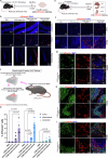
Fig. 6. Cre recombination in R26-LSL-tdTomato reporter mice by VEDIC and VFIC engineered EVs after local (ICV or osmotic pump ICV) and systemic (IP) injections.
a Workflow for the intracerebroventricular (ICV) injection of engineered EVs to deliver Cre in CNS of R26-LSL-tdTomato reporter mice. b TdTomato expression analyzed by immunofluorescence in different regions of CNS after ICV injection of engineered EVs. Scale bar, 50 µm for cerebellum and cortex, and 200 µm for hippocampus. c Workflow for the osmotic pump ICV injection of engineered EVs to transfer Cre to CNS in R26-LSL-tdTomato reporter mice. d Percentage of tdTomato+ cells in the brain tissues after osmotic pump ICV injection of engineered EVs quantified by flow cytometry, analyzed 4 days after the infusion. n = 4 mice for PBS group and n = 5 mice for other groups. e Schematic workflow for the intraperitoneal (IP) injection of engineered EVs into R26-LSL-tdTomato reporter mice. f TdTomato expression in liver and spleen after IP injection of engineered EVs, analyzed one-week post injection. Scale bar, 50 µm. g Co-staining of tdTomato and the T cell marker CD3 in spleen as detected by immunofluorescence one week after IP injection of engineered EVs. Scale bar, 50 µm. h Co-staining of tdTomato and the B cell marker B220 in spleen one week after IP injection of engineered EVs. Scale bar, 50 µm. i Co-staining of tdTomato and the macrophage marker F4/80 in spleen one week after IP injection of engineered EVs. Scale bar, 50 µm. n = 3 mice per group, representative images for (b) and (fi). Two-way ANOVA (Tukey) multiple comparisons test was used for analysis of (d). Data are shown as mean ± SD. c Created in BioRender.com, Zheng, W. (2025) https://BioRender.com/z93x284. Exact statistical analysis was reported in the Source data and Source data are provided as a Source Data file.
Fig. 7
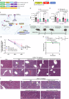
Fig. 7. Treatment of LPS-induced systemic inflammation using VEDIC and VFIC-EV mediated delivery of a super-repressor of NF-ĸB.
a, b Design of the constructs and reporter cells utilized for delivery and assessment of a super-repressor of NF-ĸB by engineered EVs. c Schematic illustration how the EV-delivered super-repressor of NF-ĸB inhibits NF-ĸB activity. d, e Luciferase activity from HEK-NF-ĸB reporter cells, 24 or 48 h after treatment with engineered EVs (TNF-α stimulation for 6 h before harvesting cells), in 24-well plates. f Schematic illustration of the workflow for the treatment of LPS-induced systemic inflammation by engineered EVs in mice (5×10¹¹ EVs/mouse per dose). g, h Percentage of body weight loss and group survival in mice after LPS and engineered EV injections. n = 10 mice per group. i Representative histology images (hematoxylin-eosin stain) of liver to show the aggregation of inflammatory cells (upper panel, yellow arrows indicate the aggregated inflammatory cells in portal areas) and the hydropic degeneration of hepatocytes (lower panel, red arrows indicate the hydropic degeneration of hepatocytes) after LPS induction. Scale bar, 50 µm. j Accumulation of inflammatory cells in the proximal region of renal tubules as shown by the representative histology images (yellow arrows indicate the inflammatory cells). Scale bar, 50 µm. Two-way (Tukey) ANOVA multiple comparisons test was used for analysis of (d, e, and g); Log-rank (Mantel-Cox) test was used for the analysis of survival curve (h). Experiments were done with 8 biological replicates for (d and e) and data are shown as mean ± SD. c, f Created in BioRender.com, Zheng, W. (2025) https://BioRender.com/s30x956 and Zheng, W. (2025) https://BioRender.com/j46d117 respectively. Exact statistical analysis was reported in the Source data and Source data are provided as a Source Data file.

References

    1. Messersmith, W. A. & Ahnen, D. J. Targeting EGFR in Colorectal Cancer. N. Engl. J. Med.359, 1834–1836 (2022). - PubMed
    1. Reck, M. et al. Safety and patient-reported outcomes of atezolizumab plus chemotherapy with or without bevacizumab versus bevacizumab plus chemotherapy in non-small-cell lung cancer. J. Clin. Oncol.38, 2530–2542 (2020). - PMC - PubMed
    1. Stewart, M. P. et al. In vitro and ex vivo strategies for intracellular delivery. Nature538, 183–192 (2016). - PubMed
    1. Goswami, R., Jeon, T., Nagaraj, H., Zhai, S. & Rotello, V. M. Accessing Intracellular Targets through Nanocarrier-Mediated Cytosolic Protein Delivery. Trends Pharm. Sci.41, 743–754 (2020). - PMC - PubMed
    1. D’Astolfo, D. S. et al. Efficient intracellular delivery of native proteins. Cell161, 674–690 (2015). - PubMed

LinkOut - more resources