. 2025 Apr 29;15(1):15022.
doi: 10.1038/s41598-025-99979-6.

BMP-2 mRNA-transfected BMSCs promote superior calvarial bone regeneration

Affiliations

BMP-2 mRNA-transfected BMSCs promote superior calvarial bone regeneration

Theeraphat Surisaeng et al. Sci Rep. .

Abstract

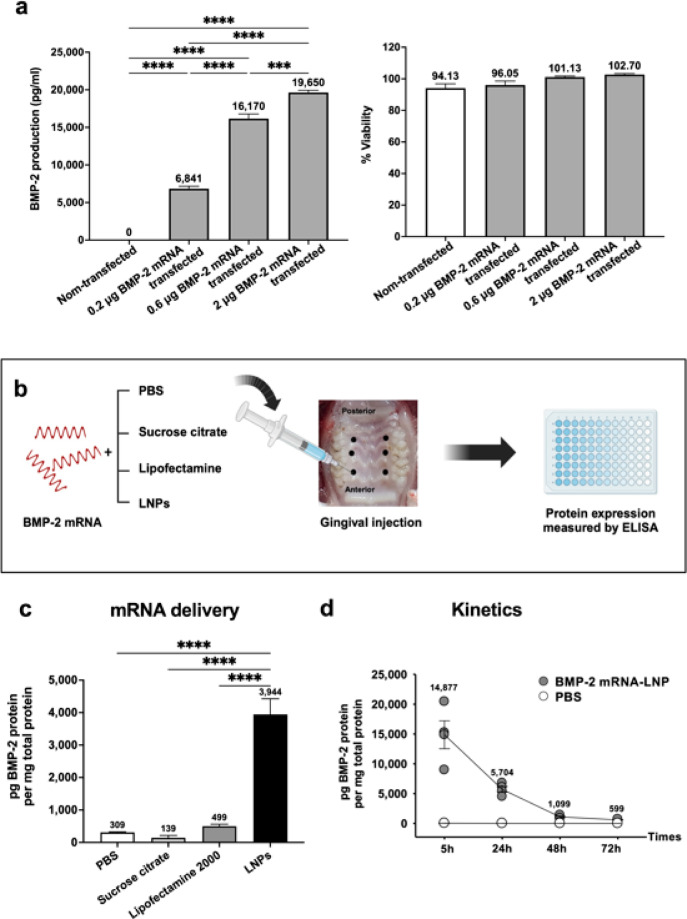
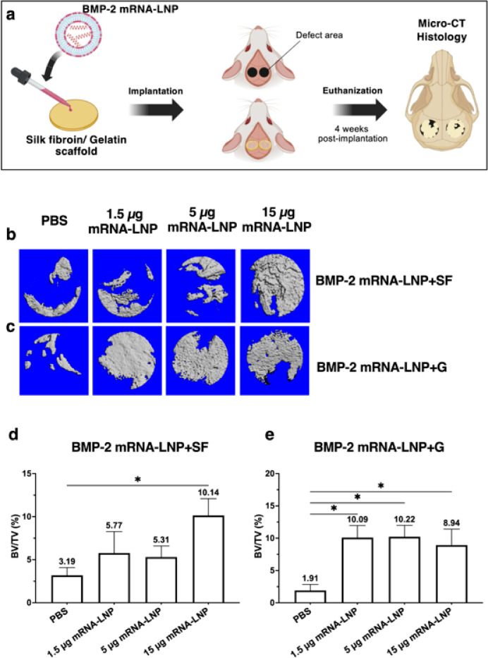
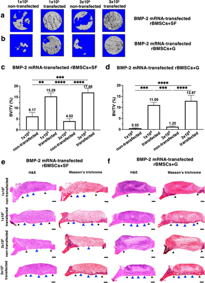
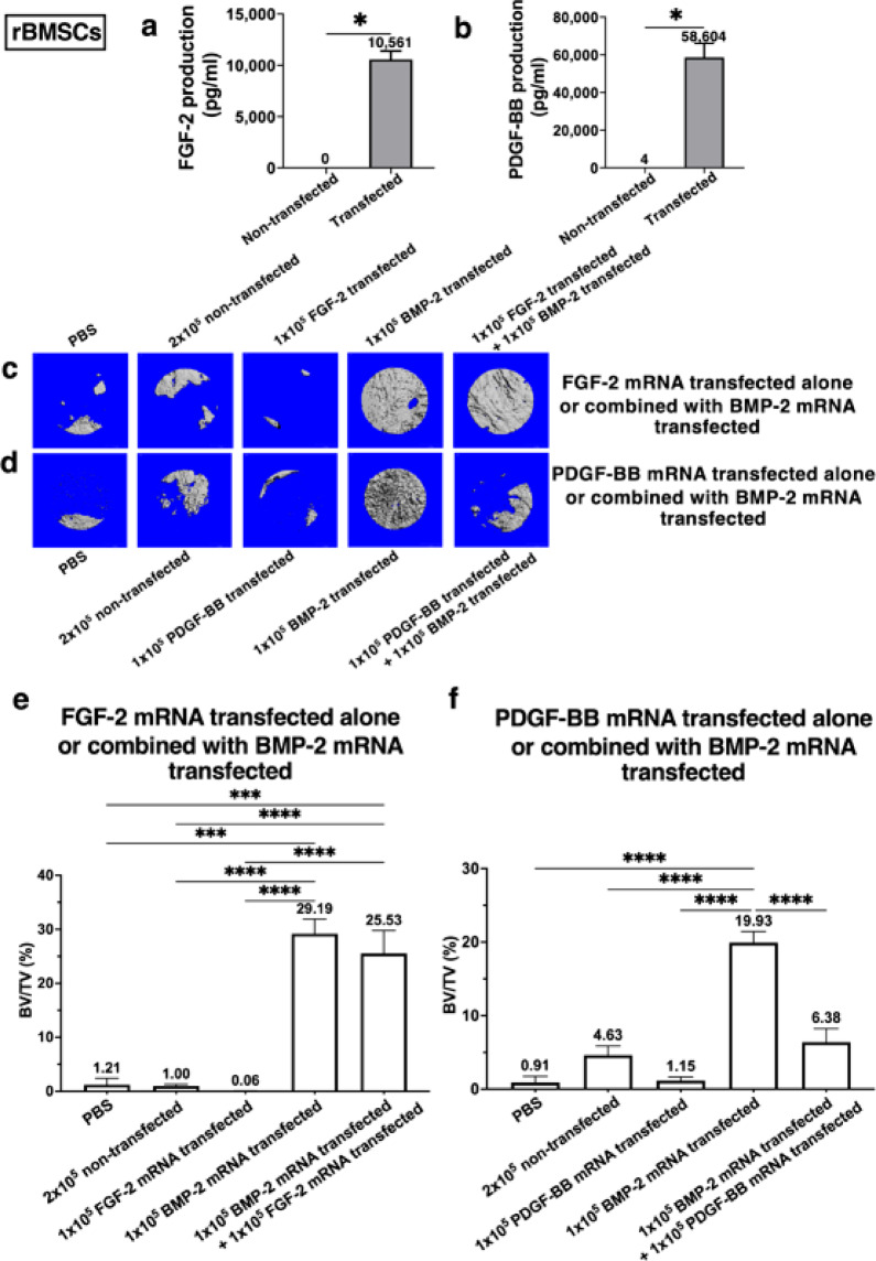
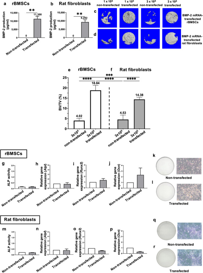
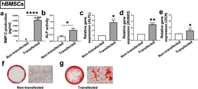
Large critical-size bone defects in the oral and craniofacial region are difficult to regenerate. We evaluated the effectiveness of mRNA encoding bone morphogenic protein-2 (BMP-2) in enhancing bone regeneration using a rat calvarial defect model. Two delivery approaches were investigated: (1) in vivo application of BMP-2 mRNA encapsulated in lipid nanoparticles incorporated in a scaffold, and (2) application of ex vivo BMP-2 mRNA-transfected rat bone marrow mesenchymal stem cells (rBMSCs), loaded on a scaffold and implanted into calvarial defects. The direct application of BMP-2 mRNA encapsulated in lipid nanoparticles improved bone regeneration as indicated by micro-computed tomography analysis. The enhancement was even more pronounced with ex vivo transfected rBMSCs. rBMSCs transfected with FGF-2 mRNA did not improve bone regeneration, either alone or combined with BMP-2 mRNA-transfected rBMSCs. Similarly, PDGF-BB mRNA-transfected rBMSCs failed to enhance bone regeneration alone and notably suppressed BMP-2 mRNA-transfected rBMSCs' effects. Interestingly, BMP-2 mRNA-transfected rat fibroblasts showed comparable bone regeneration to transfected rBMSCs. Osteogenic differentiation was absent in BMP-2 mRNA-transfected rBMSCs, implying that they may primarily serve as a source of translated BMP-2 for bone regeneration rather than undergoing osteogenic differentiation. These findings highlight the translational potential of BMP-2 mRNA for bone regeneration, particularly in oral and craniofacial applications.


Conflict of interest statement

Declarations. Competing interests: The authors declare no competing interests.

Figures

Fig. 1
Fig. 1
Optimization of modified BMP-2 mRNA delivery system. (a) Transfection of HEK 293T cells with BMP-2 mRNA complexed with Lipofectamine 2000 resulted in BMP-2 expression without cytotoxicity. Data are mean ± SE of 3 separated experiments; ***P < 0.001, ****P < 0.0001; one-way ANOVA followed by Tukey’s test. (b) Schematic view of the in vivo experimental process. Rat gingiva (palate) was injected with 30 μg of BMP-2 mRNA formulated with various vehicles, including PBS, sucrose citrate buffer, Lipofectamine 2000, and LNPs (5 μg of mRNA in 5 μl per site, with a total of 6 injection sites). (c) After 24 h, the whole gingival tissues were harvested, homogenized, and assessed for BMP-2 production using ELISA. Data are mean ± SE; n = 3 animals; ****P < 0.0001; one-way ANOVA followed by Tukey’s test. (d) Time course of BMP-2 production in rat gingiva after injection with BMP-2 mRNA-LNP. Each symbol represents one animal. Data are mean ± SE; n = 4 animals.
Fig. 2
Fig. 2
In vivo delivery of BMP-2 mRNA-LNP enhanced bone regeneration in rat calvarial defects. (a) Schematic view of the experimental process. (b,c) Representative μCT images of rat calvarial bone regeneration after 4-week implantation with BMP-2 mRNA-LNP-loaded SF scaffolds (b) or G scaffolds (c), in comparison to PBS-loaded scaffold controls. (d,e) Quantitative analysis of % bone volume (BV/TV) in defects treated with BMP-2 mRNA-LNP-loaded SF scaffolds (d) or G scaffolds (e), compared to PBS loaded scaffold controls. Data are mean ± SE; n =10 defects; *P < 0.05; one-way ANOVA followed by Dunnett’s test. SF: silk fibroin, G: gelatin-silk fibroin composite.
Fig. 3
Fig. 3
Superior improvement in calvarial bone regeneration using ex vivo BMP-2 mRNA-transfected rBMSCs. (a,b) Representative μCT images of rat calvarial bone regeneration with BMP-2 mRNA-transfected rBMSCs, loaded into SF scaffolds (a) or G scaffolds (b) compared to the non-transfected rBMSC controls. (c,d) Quantitative analysis of % bone volume (BV/TV) in defects treated with BMP-2 mRNA-transfected rBMSCs, loaded into SF scaffolds (c) or G scaffolds (d), compared to the non-transfected rBMSC controls. Data are mean ± SE; n = 10 defects; **P < 0.01, ***P < 0.001, ****P < 0.0001; one-way ANOVA followed by Tukey’s test. Histological examination confirmed bone formation in the defects. (e,f) Representative images of hematoxylin and eosin staining and Masson’s trichrome staining (n = 4 defects) provide an overview of defects treated with BMP-2 mRNA-transfected rBMSCs and non-transfected rBMSCs, loaded onto SF scaffolds (e) and G scaffolds (f). Scale bars = 500 μm. black filled triangle: host bone, blue filled triangle: new bone. SF: silk fibroin, G: gelatin-silk fibroin composite.
Fig. 4
Fig. 4
The effect of FGF-2 mRNA-transfected rBMSCs or PDGF-BB mRNA-transfected rBMSCs on calvarial bone regeneration. (a,b) In vitro transfection of rBMSCs with FGF-2 mRNA (a) or PDGF-BB mRNA (b) resulted in the production of corresponding translated proteins in the culture supernatant 24 h post-transfection. Data are mean ± SE; n = 4; *P < 0.05; Mann-Whitney U test. c, d Representative μCT images of rat calvarial bone regeneration with FGF-2 mRNA-transfected rBMSC-loaded scaffolds, both alone and in combination with BMP-2 mRNA-transfected rBMSCs (c), as well as with PDGF-BB mRNA- transfected rBMSC-loaded scaffolds alone and in combination with BMP-2 mRNA-transfected rBMSCs (d). (e,f) Quantitative analysis of % bone volume (BV/TV) in defects treated with FGF-2 mRNA-transfected BMSC-loaded scaffolds alone and in combination with BMP-2 mRNA- transfected rBMSCs (e) or with PDGF-BB mRNA-transfected BMSCs alone and in combination with BMP-2 mRNA-transfected rBMSCs (f). Data are mean ± SE; n = 6 defects; ***P < 0.001, ****P < 0.0001; one-way ANOVA followed by Tukey’s test.
Fig. 5
Fig. 5
Comparison of the efficacy in calvarial bone regeneration between BMP-2 mRNA-transfected rBMSCs and BMP-2 mRNA-transfected rat fibroblasts. In vitro transfection of BMP-2 mRNA in rBMSCs (a) or rat fibroblasts (b) resulted in the production of translated BMP-2 in the culture supernatants 24 h post-transfection. Data are mean ± SE of 3 separated experiments; **P < 0.01; Mann-Whitney U test. (c,d) Representative μCT images of rat calvarial bone regeneration with BMP-2 mRNA-transfected rBMSCs (c) and BMP-2 mRNA-transfected rat fibroblasts (d). (e,f) Quantitative analysis of % bone volume (BV/TV) in defects treated with BMP2-mRNA-transfected rBMSCs (e) or with BMP-2 mRNA- transfected rat fibroblasts (f). Data are mean ± SE; n = 6 defects; ***P < 0.001, ****P < 0.0001; one-way ANOVA followed by Tukey’s test. g-l In vitro osteogenic differentiation of BMP-2 mRNA-transfected rBMSCs; ALP activity (g), gene expression of Alpl (h), Runx2 (i), and Ocn (j). Data are mean ± SE of 3 separated experiments. A representative of Alizarin Red staining for mineralization deposits in non-transfected cells (k), and transfected cells (l). m-r In vitro osteogenic differentiation of BMP-2 mRNA-transfected rat fibroblasts; ALP activity (m), gene expression of Alpl (n), Runx (o), and Ocn (p). A representative of Alizarin Red staining for mineralization deposits in non-transfected cells (q), and transfected cells (r).
Fig. 6
Fig. 6
In vitro protein expression and osteogenic differentiation of BMP-2 mRNA-transfected hBMSCs. (a) In vitro transfection with BMP-2 mRNA in hBMSCs resulted in the production of translated BMP-2 in the culture supernatant 24 h post-transfection. Data are mean ± SE of 3 separated experiments; ****P < 0.001; Mann-Whitney U test. (bg) Osteogenic differentiation of BMP-2 mRNA-transfected hBMSCs; ALP activity (b). Data are mean ± SE of 3 separated experiments; *P < 0.05; Mann-Whitney U test. Gene expression of ALPL (c), RUNX2 (d), and OCN (e). Data are mean ± SE of 3 separated experiments; *P < 0.05, **P < 0.01; one-sample t-test. Representative images of Alizarin Red staining for mineralization deposits in non-transfected cells (f), and transfected cells (g).

Similar articles

Cited by

References

    1. Dimitriou, R., Jones, E., McGonagle, D. & Giannoudis, P. V. Bone regeneration: current concepts and future directions. BMC Med.9, 66 (2011). - PMC - PubMed
    1. Venkataiah, V. S. et al. Clinical applications of cell-scaffold constructs for bone regeneration therapy. Cells10, 2687 (2021). - PMC - PubMed
    1. Wikesjö, U. M., Huang, Y. H., Polimeni, G. & Qahash, M. Bone morphogenetic proteins: a realistic alternative to bone grafting for alveolar reconstruction. Oral Maxillofac. Surg. Clin. North. Am.19, 535–551, vi-vii (2007). - PubMed
    1. Watson-Levings, R. S. et al. Gene therapy in orthopaedics: progress and challenges in pre-clinical development and translation. Front. Bioeng. Biotechnol.10, 901317 (2022). - PMC - PubMed
    1. Fiorellini, J. P. et al. Randomized study evaluating recombinant human bone morphogenetic protein-2 for extraction socket augmentation. J. Periodontol. 76, 605–613 (2005). - PubMed

LinkOut - more resources