Prostate microcalcification crystallography as a marker of pathology
- PMID: 40301587
- PMCID: PMC12041448
- DOI: 10.1038/s41598-025-98692-8
Prostate microcalcification crystallography as a marker of pathology
Abstract
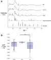
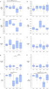
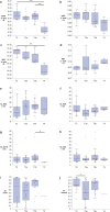
Prostate cancer remains the most common male cancer; however, treatment regimens remain unclear in some cases due to a lack of agreement in current testing methods. Therefore, there is an increasing need to identify novel biomarkers to better counsel patients about their treatment options. Microcalcifications offer one such avenue of exploration. Microfocus spectroscopy at the i18 beamline at Diamond Light Source was utilised to measure X-ray diffraction and fluorescence maps of calcifications in 10 µm thick formalin fixed paraffin embedded prostate sections. Calcifications predominantly consisted of hydroxyapatite (HAP) and whitlockite (WH). Kendall's Tau statistics showed weak correlations of 'a' and 'c' lattice parameters in HAP with GG (rτ = - 0.323, p = 3.43 × 10⁻⁴ and rτ = 0.227, p = 0.011 respectively), and a negative correlation of relative zinc levels in soft tissue (rτ = - 0.240, p = 0.022) with GG. Negative correlations of the HAP 'a' axis (rτ = - 0.284, p = 2.17 × 10⁻³) and WH 'c' axis (rτ = - 0.543, p = 2.83 × 10⁻⁴) with pathological stage were also demonstrated. Prostate calcification chemistry has been revealed for the first time to correlate with clinical markers, highlighting the potential of calcifications as biomarkers of prostate cancer.
Keywords: Biomarkers; Calcification; Prostate cancer.
© 2025. The Author(s).
Conflict of interest statement
Declarations. Ethics approval and consent to participate: The project was approved by the NRES Committee North West—Haydock (Ref: 25/NW/0013) under the Research Tissue Bank ethical approval of the Human Biomaterials Resource Centre, and all analyses were conducted in accordance with relevant regulations. Informed Consent was collected from all the individuals and is held by the Human Biomaterials Resource Centre for the samples analysed. The study was conducted in accordance with the Declaration of Helsinki. Competing interests: The authors declare no competing interests.
Figures


Similar articles
-
Stromal microcalcification in prostate.Malays J Pathol. 2001 Jun;23(1):31-3. Malays J Pathol. 2001. PMID: 16329545
-
A multi-modal exploration of heterogeneous physico-chemical properties of DCIS breast microcalcifications.Analyst. 2022 Apr 11;147(8):1641-1654. doi: 10.1039/d1an01548f. Analyst. 2022. PMID: 35311860 Free PMC article.
-
Association between asymptomatic inflammatory prostatitis NIH category IV and prostatic calcification in patients with obstructive benign prostatic hyperplasia.Minerva Urol Nefrol. 2016 Jun;68(3):242-9. Epub 2015 May 27. Minerva Urol Nefrol. 2016. PMID: 26013949
-
Microcalcifications in breast cancer: Lessons from physiological mineralization.Bone. 2013 Apr;53(2):437-50. doi: 10.1016/j.bone.2013.01.013. Epub 2013 Jan 18. Bone. 2013. PMID: 23334083 Review.
-
Structural and molecular biology of PSP94: Its significance in prostate pathophysiology.Front Biosci (Landmark Ed). 2018 Jan 1;23(3):535-562. doi: 10.2741/4604. Front Biosci (Landmark Ed). 2018. PMID: 28930560 Review.
References
-
- Cancer Research UK. Prostate Cancer Statistics. https://www.cancerresearchuk.org/health-professional/cancer-statistics/s... (2024).
MeSH terms
Substances
Grants and funding
LinkOut - more resources
Full Text Sources
Medical

