

Vision transformer-based diagnosis of lumbar disc herniation with grad-CAM interpretability in CT imaging
- PMID: 40301802
- PMCID: PMC12039304
- DOI: 10.1186/s12891-025-08602-2
Vision transformer-based diagnosis of lumbar disc herniation with grad-CAM interpretability in CT imaging
Abstract
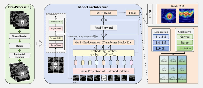
Background: In this study, a computed tomography (CT)-vision transformer (ViT) framework for diagnosing lumbar disc herniation (LDH) was proposed for the first time by taking advantage of the multidirectional advantages of CT and a ViT.
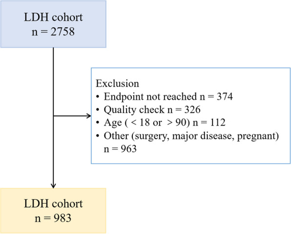
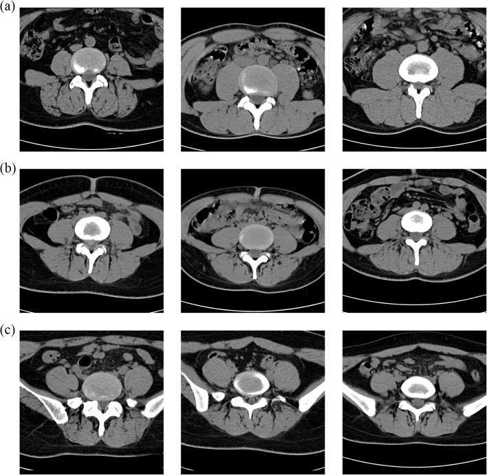
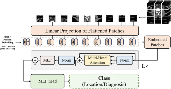
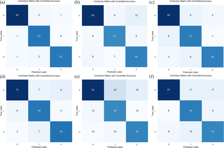
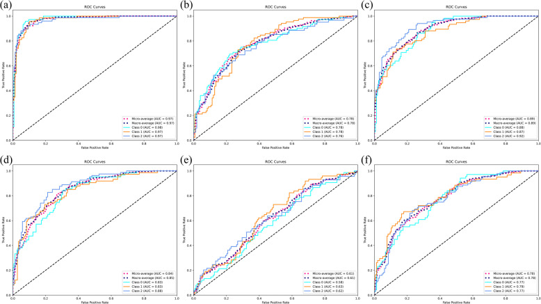
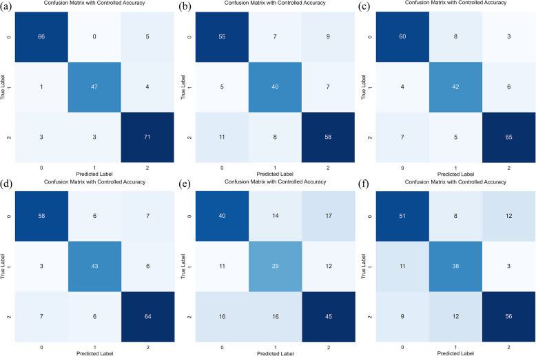
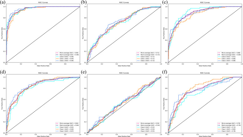
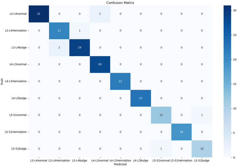
Methods: The proposed ViT model was trained and validated on a dataset consisting of 983 patients, including 2100 CT images. We compared the performance of the ViT model with that of several convolutional neural networks (CNNs), including ResNet18, ResNet50, LeNet, AlexNet, and VGG16, across two primary tasks: vertebra localization and disc abnormality classification.
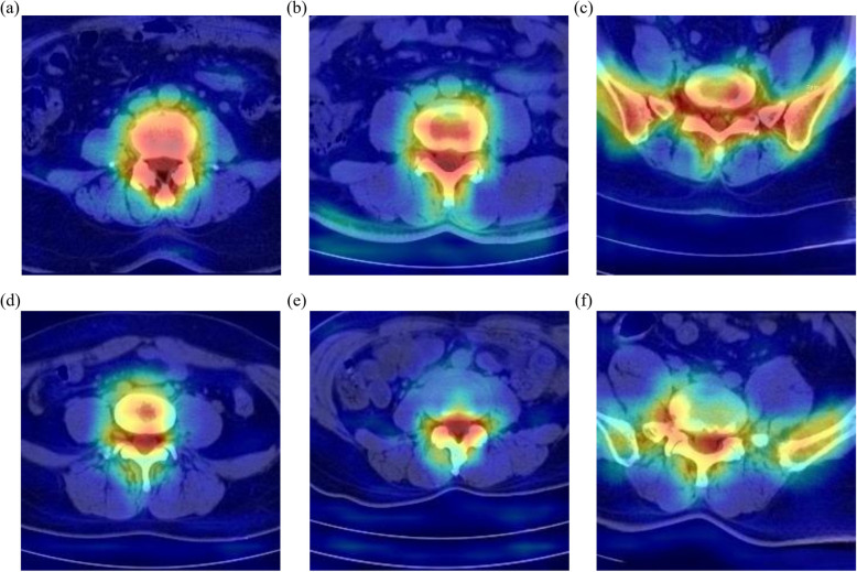
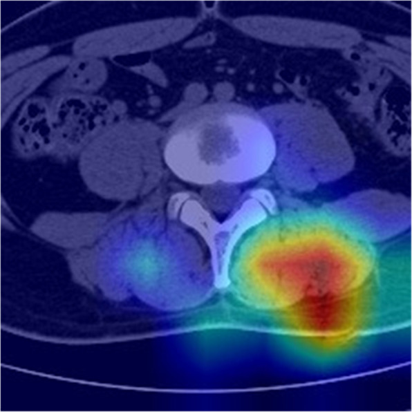
Results: The integration of a ViT with CT imaging allowed the constructed model to capture the complex spatial relationships and global dependencies within scans, outperforming CNN models and achieving accuracies of 97.13% and 93.63% in terms of vertebra localization and disc abnormality classification, respectively. The performance of the model was further validated via gradient-weighted class activation mapping (Grad-CAM), providing interpretable insights into the regions of the CT scans that contributed to the model predictions.
Conclusion: This study demonstrated the potential of a ViT for diagnosing LDH using CT imaging. The results highlight the promising clinical applications of this approach, particularly for enhancing the diagnostic efficiency and transparency of medical AI systems.
Keywords: CT; Deep learning; Diagnostic accuracy; Grad-CAM; LDH; Medical imaging; ViT.
© 2025. The Author(s).
Conflict of interest statement
Declarations. Ethics approval and consent to participate: Our study adhered to the Declaration of Helsinki. This study received approval from the Ethics Committee of the First Affiliated Hospital of Anhui University of Traditional Chinese Medicine (no. 2024MCZQ28), and the ethics committee waived the need to consent to participate because of the minimal risk involved. Consent for publication: Not applicable. Competing interests: The authors declare no competing interests.
Figures
References
MeSH terms
Grants and funding
LinkOut - more resources
Full Text Sources
Medical
