. 2025 Apr 29;22(1):125.
doi: 10.1186/s12974-025-03455-x.

Repurposing of epalrestat for neuroprotection in parkinson's disease via activation of the KEAP1/Nrf2 pathway

Affiliations

Repurposing of epalrestat for neuroprotection in parkinson's disease via activation of the KEAP1/Nrf2 pathway

Huafang Jia et al. J Neuroinflammation. .

Abstract

Background: Epalrestat (EPS), an aldose reductase inhibitor, is used to alleviate peripheral nerve disorder of diabetic patients in clinical therapy. Even though EPS exerted effects in central nervous system diseases, the neuroprotection and underlying molecular mechanism in neurodegenerative diseases, especially Parkinson's disease (PD), remains obscure. Our study aimed to investigate the potential of EPS suppressed PD progression both in vivo and in vitro.

Methods: We used 1-methyl-4-phenylpyridillium ion (MPP+)-treated PD cells and 1-methyl-4-phenyl-1,2,3,6-tetrahydropyridine (MPTP)-treated PD mice to investigate the protective function and molecular mechanism of EPS in PD. EPS was administered three times daily through oral route 3 days before model establishment for 5 consecutive days. Behavioral manifestation of mice was conducted using open field test, rotarod test and CatWalk gait analysis. Immunofluorescence was used to detect dopaminergic (DAergic) neurons survival in the substantia nigra. Subsequently, oxidative stress, mitochondrial function and KEAP1/Nrf2 signaling pathway in PD models were detected through molecular biology methods to assess the effect and downstream mechanisms of EPS on PD. Molecular docking, surface plasmon resonance and cellular thermal shift assay were used to verify the direct binding of EPS and KEAP1.

Results: We found that EPS exhibited potent antiparkinsonian activity in PD models both in vivo and in vitro. PD models treated with EPS manifested alleviated oxidative stress and mitochondrial dysfunction. Furthermore, we found EPS activated the Nrf2 signaling pathway which contributed to DAergic neurons survival in PD models. Particularly, we firstly confirmed that EPS competitively binds to KEAP1 and enhanced its degradation, thereby activating the Nrf2 signaling pathway.

Conclusions: Collectively, EPS attenuates oxidative stress and mitochondrial dysfunction by directly binding KEAP1 to activate the KEAP1/Nrf2 signaling pathway, further reducing DAergic neurons damage. These findings suggest that EPS has great potential to become a therapeutic for PD as a clinically effective and safe medicine.

Keywords: Epalrestat; KEAP1; Mitochondrial dysfunction; Nrf2; Oxidative stress; Parkinson’s disease.


Conflict of interest statement

Declarations. Ethics approval and consent to participate: All animal experiments were performed following international guidelines and conducted in accordance with the Animal Ethics Committee of Qingdao University (QDU-AEC-2024766). Consent for publication: Not applicable. Competing interests: The authors declare no competing interests.

Figures
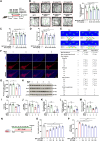
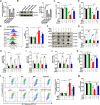
Fig. 1
Fig. 1
EPS improved motor function and attenuated the injury of DAergic neurons in MPP+/MPTP-treated PD model. A Schematic illustration in mice experiment. Mice were pretreated with EPS (25 mg/kg, 50 mg/kg and 100 mg/kg) for 3 days followed by MPTP (25 mg/kg) for 5 days. (B) OFT. n = 6. C Rotarod test. n = 6. D Pole test. n = 6. E CatWalk gait analysis. n = 8. Data shows: no significance observed (↔), significant increase observed (↑) and significant reduction observed (↓).F, G Representative images of TH+ neurons in the SNpc. n = 8. H-J Representative blots and analysis for TH, Bcl-2 and Bax were detected by specific antibodies in the SN. n = 8, 12. (K-M) DA, DOPAC and HVA content were measured by HPLC-ECD. n = 12. N Schematic illustration in cell experiment. N2a cells were pretreated with EPS (100 μM) for 4 h followed by MPP+ (100 μM) for 24 h. O CCK-8 experiment was used to detect cell viability. P Cytotoxicity was measured by LDH release. Data are expressed as mean ± SEM. *P < 0.05, **P < 0.01, ***P < 0.001, versus the indicated group. #P < 0.05, ##P < 0.01, ###P < 0.001, versus MPTP group. Ctrl, control; ns, no significance
Fig. 2
Fig. 2
EPS alleviates oxidative stress and mitochondrial dysfunction. A ROS was measured by flow cytometry using C11-BODIPY581/591 in N2a cells. B-E, G-I SOD, CAT, GSH and NADPH levels were detected in N2a cells and the SN of mice. F MDA levels detected in the SN of mice represent lipid peroxidation. J Δψm was detected using JC-1 by flow cytometry in N2a cells. K, O The ultrastructure of mitochondria (the position indicated by the red arrow) in N2a cells and DAergic neurons were observed by TEM. L, M Mitochondrial ROS and intracellular calcium concentration were measured using mitoSOX and Fluo-4 AM by flow cytometry in N2a cells. N, P ATP levels were detected in N2a cells and the SN of mice. Data are expressed as mean ± SEM. *P < 0.05, **P < 0.01, ***P < 0.001 versus the indicated group. Ctrl, control
Fig. 3
Fig. 3
Network pharmacology analysis of EPS on PD. A Venn diagram of the common target of EPS and PD. B PPI network of the top 30 hub overlapping genes with high degree scores. C Nrf2 pathway for GSEA enrichment analysis on the GSE26927 database. D The mRNA transcription levels of KEAP1, NFE2L2, GCLC, HMOX1 and NQO1 in the PD patients’ groups of GSE26927. E The mRNA transcription levels of keap1, nfe2l2, gclm, hmox1 and nqo1 in the animal model groups of GSE30. Data are expressed as mean ± SEM. *P < 0.05, **P < 0.01, ***P < 0.001 versus the indicated group. Ctrl, control
Fig. 4
Fig. 4
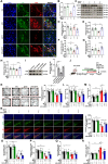
The direct binding interactions between EPS and KEAP1. A Molecular docking between EPS and KEAP1. B SPR analysis was used to verify the interaction between EPS and KEAP1. C CETSA for interaction between EPS and KEAP1. D Degradation of KEAP1 protein by EPS when CHX inhibits protein synthesis in N2a cells. (E-J) CETSA for interaction between EPS and KEAP1 mutant (S363A, A414A, S555A, S602A). (K) CCK-8 experiment was used to detect cell viability. L, M ROS was measured by flow cytometry using C11-BODIPY581/591. N Δψm was detected using JC-1 by flow cytometry. Data are expressed as mean ± SEM. *P < 0.05, **P < 0.01, ***P < 0.001 versus the indicated group. n = 3. Ctrl, control
Fig. 5
Fig. 5
EPS binds KEAP1 promotes the activation of Nrf2 signaling pathway. A, B Representative images of Nrf2 and DAPI. The fluorescence of Nrf2 in the nucleus was analyzed in columns graph (n = 110–130 cells for each group). C-E Nucleus and plasma proteins were isolated. Representative blots and analysis for Nrf2 were detected by specific antibodies. FH GCLC, HO-1 and NQO1 mRNA levels were measured by using RT-qPCR. I-N Representative blots and analysis for KEAP1, Nrf2, GCLC, HO-1 and NQO1 were detected by specific antibodies. Data are expressed as mean ± SEM. *P < 0.05, **P < 0.01, ***P < 0.001 versus the indicated group. n = 3. Ctrl, control; A.U., arbitrary units
Fig. 6
Fig. 6
Knockdown of Nrf2 neutralized EPS mediated antioxidant and mitochondrial protective effect in MPP+-treated N2a cells. A Nrf2 mRNA levels were measured using RT-qPCR. B Representative blots and analysis for Nrf2 detected by specific antibodies in Nrf2 knockdown cells. C, D Cell viability was measured by CCK-8 after using ML385 or knockdown of Nrf2. E ROS was measured by flow cytometry using C11-BODIPY581/591 in Nrf2 knockdown cells. F-J Representative blots and analysis for Nrf2, GCLC, HO-1 and NQO1 detected by specific antibodies in Nrf2 knockdown cells. K GSH level were detected using ELISA in Nrf2 knockdown cells. L Δψm was detected using JC-1 by flow cytometry in Nrf2 knockdown cells. M ATP levels were detected in Nrf2 knockdown cells. Data are expressed as mean ± SEM. *P < 0.05, **P < 0.01, ***P < 0.001 versus the indicated group. n = 3. Ctrl, control; NC, negative control
Fig. 7
Fig. 7
Knockdown of Nrf2 attenuated EPS mediated antioxidant capacity and the curative activity. A, B Representative images of Nrf2, TH and DAPI. The fluorescence of Nrf2 in the nucleus was analyzed in columns graph (n = 6, 20–40 cells for each group). C-H Representative blots and analysis for KEAP1, Nrf2, GCLC, HO-1 and NQO1 were detected by specific antibodies in the SN of mice. n = 12. I Representative blots and analysis for Nrf2 were detected by specific antibodies in the SN of mice injected with AAV-siNrf2. n = 6. J Schematic illustration in mice experiment. Mice were injected with AAV-siNrf2. After 21 Days, mice were pretreated with EPS (50 mg/kg) for 3 days followed by MPTP (25 mg/kg) for 5 days. K OFT. n = 12. L Rotarod test. n = 12. (M) Pole test. n = 12. N Representative images of TH+ cells in the SNpc. n = 8. O-Q DA, DOPAC and HVA levels in the striatum of mice were measured by HPLC-ECD. n = 12. (R) MDA levels were detected in the SN of mice. n = 8. Data are expressed as mean ± SEM. *P < 0.05, **P < 0.01, ***P < 0.001 versus the indicated group. Ctrl, control; NC, negative control; A.U., arbitrary units
Fig. 8
Fig. 8
The antioxidant and mitochondrial protective effects of EPS in PD. EPS binds to KEAP1, which further promotes the activation of Nrf2 signaling pathway, thereby suppressing oxidative stress and enhancing mitochondrial function, ultimately protecting DAergic neurons in PD

Similar articles

References

    1. Berg D, Postuma RB, Bloem B, Chan P, Dubois B, Gasser T, Goetz CG, Halliday GM, Hardy J, Lang AE, et al. Time to redefine PD? Introductory statement of the MDS Task Force on the definition of Parkinson’s disease. Mov Disord. 2014;29:454–62. - PMC - PubMed
    1. Xu T, Dong W, Liu J, Yin P, Wang Z, Zhang L, Zhou M. Disease burden of Parkinson’s disease in China and its provinces from 1990 to 2021: findings from the global burden of disease study 2021. Lancet Reg Health West Pac. 2024;46: 101078. - PMC - PubMed
    1. Elkouzi A, Vedam-Mai V, Eisinger RS, Okun MS. Emerging therapies in Parkinson disease - repurposed drugs and new approaches. Nat Rev Neurol. 2019;15:204–23. - PMC - PubMed
    1. Choudhary S, Silakari O. Virtual screening of epalrestat mimicking selective ALR2 inhibitors from natural product database: auto pharmacophore, ADMET prediction and molecular dynamics approach. J Biomol Struct Dyn. 2022;40:6052–70. - PubMed
    1. He J, Gao HX, Yang N, Zhu XD, Sun RB, Xie Y, Zeng CH, Zhang JW, Wang JK, Ding F, et al. The aldose reductase inhibitor epalrestat exerts nephritic protection on diabetic nephropathy in db/db mice through metabolic modulation. Acta Pharmacol Sin. 2019;40:86–97. - PMC - PubMed

MeSH terms

LinkOut - more resources