. 2025 Apr 29;16(1):29.
doi: 10.1186/s13293-025-00711-w.

ERβ mediates sex-specific protection in the App-NL-G-F mouse model of Alzheimer's disease

Affiliations

ERβ mediates sex-specific protection in the App-NL-G-F mouse model of Alzheimer's disease

Aphrodite Demetriou et al. Biol Sex Differ. .

Abstract

Background: Menopausal loss of neuroprotective estrogen is thought to contribute to the sex differences in Alzheimer's disease (AD). Activation of estrogen receptor beta (ERβ) can be clinically relevant since it avoids the adverse systemic effects of ERα activation. However, very few studies have explored ERβ-mediated neuroprotection in AD, and no information on its contribution to the sex differences in AD exists. In the present study, we specifically explored the role of ERβ in mediating sex-specific protection against AD pathology in the AppNL-G-F knock-in mouse model of amyloidosis, and if surgical menopause (ovariectomy) modulates pathology in this model.

Methods: We treated male and female AppNL-G-F knock-in mice with the clinically relevant and selective ERβ agonist LY500307. A subset of the females was ovariectomized prior to treatment. Y-maze and contextual fear conditioning tests were used to assess memory performance, and biochemical assays such as qPCR, immunohistochemistry, Western blot, and multiplex immunoassays, were used to evaluate amyloid pathology.

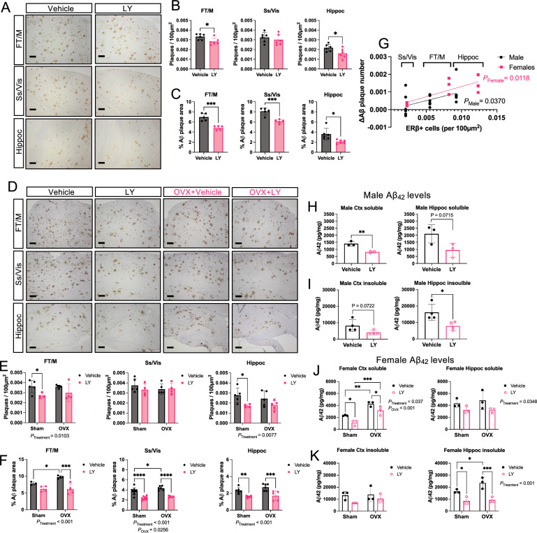
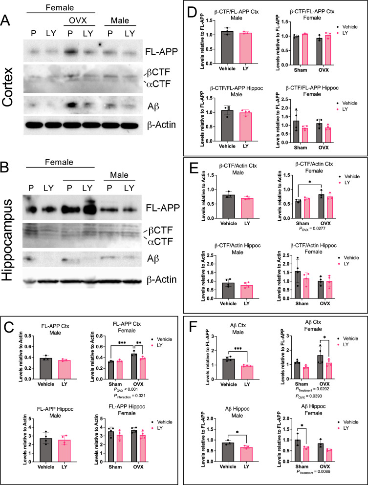
Results: We found that Female AppNL-G-F mice had higher soluble Aβ levels in cortex and hippocampus than males and more activated microglia. ERβ activation protected against amyloid pathology and cognitive decline in both male and female AppNL-G-F mice. Although ovariectomy increased soluble amyloid beta (Aβ) in cortex and insoluble Aβ in hippocampus, as well as sustained neuroinflammation after ERβ activation, it had otherwise limited effects on pathology. We further identified that ERβ did not alter APP processing, but rather exerted its protection at least partly via microglia activation in a sex-specific manner.

Conclusion: Combined, we provide new understanding to the sex differences in AD by demonstrating that ERβ protects against AD pathology differently in males and females, warranting reassessment of ERβ in combating AD.

Keywords: APP knock-in; Alzheimer’s disease; Amyloidosis; Estrogen receptor beta; Microglia; Sex differences; Sex hormone.

Plain language summary

About two-thirds of all Alzheimer’s disease (AD) patients are women. Although the reason for this sex difference is likely multifaceted, sex hormones are believed to be involved. The female sex hormone estrogen is known to mediate neuroprotection and loss of estrogen during the menopausal transition is believed to be a risk factor for AD. However, there is a gap in knowledge on how estrogenic neuroprotection occurs and if this neuroprotection is similar in men and women. In the current study, we specifically focused on one estrogen receptor, ERβ, and its role in mediating protection in a clinically relevant mouse model of AD and asked if there are any differences in this protection between male and female AD mice. Such information is of importance if proposing clinical trials targeting ERβ, which unlike targeting the ubiquitous estrogen receptor alpha (ERα), is not associated with adverse systemic effects. We found that ERβ activation indeed protects against amyloid plaque buildup and cognitive impairment in both males and females. Interestingly, this neuroprotection appeared to work differently in different brain regions and affected neuroinflammation and microglia immune cell function differently in males and females. Surgical menopause (ovariectomy) increased amyloid levels, which was counteracted by ERβ activation, and sustained high neuroinflammation but had otherwise limited effect on pathology. We provide the first study comparing ERβ-mediated protection on AD pathology in males and females, highlighting important sex differences that should be considered when proposing ERβ as a target to combat AD.


Conflict of interest statement

Declarations. Ethics approval and consent to participate: All procedures were performed in accordance with approved ethical permits (ethical approval ID 407 and ID 2199–2021, Linköping’s animal ethical board). Consent for publication: Not applicable. Competing interests: The authors declare that they have no competing interests.

Figures

Fig. 1
Fig. 1
ERβ activation improves cognitive behavior in AppNL−G−F male and female mice. A Treatment regime of AppNL−G−F mice. B Representative image of Y-maze arena. C Percent Y-maze arm alterations and D total number of arm entries of male (left) and female (right) AppNL−G−F mice treated with vehicle or ERβ agonist LY500307 (LY) (n = 7–10). E Diagram showing the fear conditioning paradigm. F Percent context-associated freezing time of male (left) and female (right) AppNL−G−F mice (n = 6–9) in the contextual fear conditioning test. Cued-associated freezing time in the contextual fear conditioning test of G male and H female AppNL−G−F mice before cue (baseline) and upon cue (tone) in a different cage context (n = 6–9). Female mice were either ovariectomized (OVX) or sham operated (Sham). * P < 0.05, ** P < 0.01, *** P < 0.001. Unpaired t-test was used for males and 2-way ANOVA for females followed by uncorrected Fisher’s LSD test for multiple comparisons. Overall significant main effects of treatment or OVX are indicated
Fig. 2
Fig. 2
Less Aβ pathology in AppNL−G−F male and female mice after ERβ activation. A Immunohistochemical representation of amyloid plaques in frontal and motor cortex (FT/M), somatosensory and visual cortex (Ss/Vis) and hippocampus (Hippoc) of male AppNL−G−F mice after vehicle or LY treatment. B Quantification of number of plaques per 100 µm2 (n = 4–6) and C percent plaque area (n = 4–6) in male AppNL−G−F mice. D Similar as in A, immunohistochemical representation of amyloid plaques in different brain regions of female AppNL−G−F mice after vehicle or LY treatment. E Quantification of number of plaques per 100 µm2 (n = 4–5) and F percent plaque area (n = 4–9) in female AppNL−G−F mice. G Linear regression analysis comparing effect size from LY treatment (vehicle vs. LY) on number of Aβ plaques in relation to average number of ERβ positive cells per 100 µm2 in different brain regions of male and female mice (n = 4–6). H Soluble and (I) insoluble Aβ42 levels in male cortex (Ctx, left) and hippocampus (Hippoc, right) (n = 3–4). (J) Soluble and (K) insoluble Aβ42 levels in female cortex (left) and hippocampus (right) (n = 3). * P < 0.05, ** P < 0.01, *** P < 0.001, **** P < 0.0001. Unpaired t-test was used for males and 2-way ANOVA for females followed by uncorrected Fisher’s LSD test for multiple comparisons. Overall significant main effects of treatment or OVX are indicated. Scale bars = 100 µm
Fig. 3
Fig. 3
ERβ activation does not alter APP processing. Western blot analysis of full-length APP (FL-APP), β-CTF and α-CTF, Aβ peptide, and β-actin in A cortex and B hippocampus of female and male AppNL−G−F mice after vehicle (V) or LY treatment, as well as after sham surgery or ovariectomy (OVX in females). Quantification of C FL-APP relative to β-actin, D β-CTF relative to FL-APP, E β-CTF relative to actin, and F Aβ relative to FL-APP in male (left), and female (right), cortex (Ctx) (top), and hippocampus (bottom) (n = 3–4). * P < 0.05, ** P < 0.01, *** P < 0.001. Statistical significance was determined using unpaired t-test for males and 2-way ANOVA for females followed by uncorrected Fisher’s LSD test for multiple comparisons. Overall significant main effects of treatment or OVX are indicated
Fig. 4
Fig. 4
ERβ activation modulates microglia activation in a sex-specific manner in AppNL−G−F mice. A Representative immunofluorescence images of male AppNL−G−F hippocampus stained with the amyloid stain AmyloGlo (magenta), Iba1 (green), and CD68 (white) after vehicle or LY treatment. Yellow dotted area (left) indicates magnified region of interest (right). Arrowheads indicate microglia with lower CD68 levels. Scale bar 100 µm (left) and 50 µm (right). Quantification in male AppNL−G−F mice of B number of Iba1 cells per 100 µm2 (n = 5–6), C percent CD68 +, Iba1 + double positive cells (n = 4–5), and D percent microglia within 20 µm radius of plaque edge (n = 5–6). E Representative immunofluorescence images of female AppNL−G−F hippocampus stained with AmyloGlo (magenta), Iba1 (green), and CD68 (white) after vehicle or LY treatment, as well as after sham surgery or ovariectomy (OVX). Yellow dotted area (left) indicates magnified region of interest (right). Arrowheads indicate microglia with lower CD68 levels. Scale bar 100 µm (left) and 50 µm (right). Quantification in female AppNL−G−F mice F number of Iba1 cells per 100 µm2 (n = 4), (G percent CD68 +, Iba1 + double positive cells (n = 4), and H percent plaque-associated microglia (n = 4). * P < 0.05, *** P < 0.001. Unpaired t-test was used for males and 2-way ANOVA for females followed by uncorrected Fisher’s LSD test for multiple comparisons. Overall significant main effects of treatment are indicated
Fig. 5
Fig. 5
Microglial and proinflammatory markers are altered upon ERβ activation in a sex-specific manner. A Representative immunofluorescence image of ERβ and Iba1 co-staining (arrowheads) in WT (left) and Esr2-KO (right) male cortex (dotted rectangle: magnified area), and B in male AppNL−G−F cortex upon vehicle or LY treatment (scale bars = 50 µm). C Quantification and D comparison of percent ERβ positive microglia in male and female AppNL−G−F brains (cortex and hippocampus) upon OVX and/or LY treatment (n = 4). Expression of the proresolving microglial markers E Trem2, and F Cx3cr1 relative to housekeeping gene Rplp0 in male (left) and female (right) AppNL−G−F hippocampus after vehicle or LY treatment, as well as after sham surgery or OVX in females (n = 3–7). Multiplex ELISA analysis of the inflammatory markers G CXCL1 (KC/GRO), H IL-12p70, and I IL-10, in male (left) and female (right) APPNL−G−F hippocampus after vehicle or LY treatment, as well as after sham surgery or ovariectomy (OVX in females) (n = 4–6). * P < 0.05, ** P < 0.01, *** P < 0.001. Unpaired t-test was used for males and 2-way ANOVA for females followed by uncorrected Fisher’s LSD test for multiple comparisons. Overall significant main effects of treatment or OVX are indicated

Update of

Similar articles

Cited by

References

    1. Maioli S, Leander K, Nilsson P, Nalvarte I. Estrogen receptors and the aging brain. Essays Biochem. 2021;65(6):913–25. 10.1042/ebc20200162. - PMC - PubMed
    1. Rettberg JR, Yao J, Brinton RD. Estrogen: a master regulator of bioenergetic systems in the brain and body. Front Neuroendocrinol. 2014;35(1):8–30. 10.1016/j.yfrne.2013.08.001. - PMC - PubMed
    1. Scheyer O, Rahman A, Hristov H, Berkowitz C, Isaacson RS, Diaz-Brinton R, et al. Female Sex and Alzheimer’s Risk: the menopause connection. the journal of prevention of Alzheimer's Disease. 2018; 5(4):225–30. 10.14283/jpad.2018.34 - PMC - PubMed
    1. Rocca WA, Grossardt BR, Geda YE, Gostout BS, Bower JH, Maraganore DM, et al. Long-term risk of depressive and anxiety symptoms after early bilateral oophorectomy. Menopause. 2008;15(6):1050–9. 10.1097/gme.0b013e318174f155. - PubMed
    1. Mishra A, Wang Y, Yin F, Vitali F, Rodgers KE, Soto M, et al. A tale of two systems: Lessons learned from female mid-life aging with implications for Alzheimer’s prevention & treatment. Ageing Res Rev. 2022;74: 101542. 10.1016/j.arr.2021.101542. - PMC - PubMed

Substances

LinkOut - more resources