. 2025 Apr;21(4):e70194.
doi: 10.1002/alz.70194.

Inhibition of hippocampal mossy fiber plasticity and episodic memory by human Aβ oligomers is prevented by enhancing cAMP signaling in Alzheimer's mice

Affiliations

Inhibition of hippocampal mossy fiber plasticity and episodic memory by human Aβ oligomers is prevented by enhancing cAMP signaling in Alzheimer's mice

Shan-Xue Jin et al. Alzheimers Dement. 2025 Apr.

Abstract

Introduction: Early episodic memory impairment in Alzheimer's disease (AD) is linked to synaptic dysfunction from amyloid β-protein oligomers (oAβ), particularly affecting the dentate gyrus mossy fiber-CA3 pathway. The APPNL-G-F mouse model exhibits early deficits in mossy fiber long-term potentiation (mf-LTP).

Methods: We administered the β-adrenergic receptor agonist isoproterenol (ISO) in vivo and phosphodiesterase type 4 inhibitor GSK356278 in vitro to assess their impact on mf-LTP and contextual fear memory. Fluorescence lifetime imaging (FLIM)-Förster resonance energy transfer (FRET) microscopy was used to visualize impaired and rescued cyclic adenosine monophosphate (cAMP) signaling in dentate gyrus neurons.

Results: ISO prevented mf-LTP impairment at 3-4 mo and improved memory by 7 mo. GSK356278 inhibited mf-LTP deficits in a dose-dependent manner. ISO also reduced hyperphosphorylation of synapsin I and microgliosis.

Discussion: These findings suggest that β-AR activation and phosphodiesterase 4 (PDE4) inhibition mitigate oAβ-induced memory deficits, supporting enhanced cAMP signaling as a therapeutic target for early AD.

Highlights: Early episodic memory deficits in AD linked to oAβ-induced synaptic dysfunction. Isoproterenol and GSK356278 improve mossy fiber-LTP and fear memory deficits. FLIM-FRET shows treatments restore cAMP signaling in dentate gyrus neurons. Isoproterenol reduces synapsin I hyperphosphorylation and microgliosis. Enhancing cAMP signaling may help mitigate early memory deficits in AD.

Keywords: APPNL‐G‐F knock‐in mice; Alzheimer's disease; Aβ oligomers; FLIM‐FRET; cAMP; episodic memory; long‐term potentiation; microgliosis; mossy fiber‐CA3 pathway; β‐adrenergic signaling.


Conflict of interest statement

D.J.S. is a director of Prothena Biosciences and ad hoc consultant to Eisai and Roche. The other authors declare no competing interests. Author disclosures are available in the supporting information.

Figures
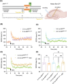
FIGURE 1
FIGURE 1
Deficits in mf‐LTP in APPNL‐G‐F mice start at age 3–4 mo, but 2 mo pretreatment with oral isoproterenol prevents this. (A) Diagram of the APP and sequence of Aβ indicating Swedish, Arctic and Iberian mutations (left), as well as schematics of slice recording (right, created with BioRender.com). Time course LTP traces show that mf‐LTP is impaired in 3–4 mo APPNL‐G‐F mice (C), but not in 2‐mo‐old APP NL‐G‐F mice (B), when compared to 3–4 mo wt mice. (D) Oral ISO prevented the defect in mf‐LTP in 3–4 mo APPNL‐G‐F mice. (E) Histograms of the average potentiation for the last 10 min of mf‐LTP recordings. Each slice used for each experiment was from a different mouse. Error bars = means ± SEMs; p‐values calculated using one‐way ANOVA with Tukey's post‐test comparing the means of recorded fEPSP slopes. Compared to the same age in wt mice, mf‐LTP is impaired in 3–4 mo APPNL‐G‐F mice (wt 156.3 ± 7.27 n = 4 vs. APPNL‐G‐F 103.1 ± 8.32; n = 9, p = 0.004) but not in 2 mo APPNL‐G‐F mice (wt 163.3 ± 13.32 n = 5 vs. APPNL‐G‐F 143 ± 4.67; n = 4, p = 0.55). ISO prevented the decrease in mf‐LTP in 3 mo APPNL‐G‐F mice (Ctrl: 103.1 ± 8.32 n = 9 vs. ISO: 157.8 ± 6.84 n = 7; p = 0.0005).
FIGURE 2
FIGURE 2
Dose‐dependence of PDE4 inhibitor GSK356278 rescue of mf‐LTP inhibition in 3–4 or 6–7 mo APPNL‐G‐F mice. GSK356278 (1 µM but not 0.1 µM) prevented the inhibition of LTP in 3–4 mo (A, C 0 µM: 112.5 ± 6.98 n = 7 vs. 0.1 µM: 104.8 ± 8.15 n = 5, p = 0.978; 0 µM: 112.5 ± 6.98 n = 7 vs. 1 µM: 172.5 ± 15.09 n = 4, p = 0.0002) or 6–7 mo (B, C 0 µM: 107.3 ± 3.39 n = 7 vs. 0.1 µM: 96.98 ± 3.53 n = 5, p = 0.926; 0 µM: 107.3 ± 3.39 n = 7 vs. 1 µM: 173.3 ± 10.61 n = 5, p < 0.0001) APPNL‐G‐F mice. Each slice used for each treatment was from a different mouse. Error bars = means ± SEMs. p‐Values calculated via one‐way ANOVA test; ***p < 0.001, ****p < 0.0001, ns, p > 0.05.
FIGURE 3
FIGURE 3
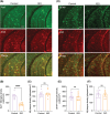
Isoproterenol prevents hyperphosphorylation of synapsin I at S9 in CA3 at 6–7 mo APPNL‐G‐F mice. (A) Schematic depiction of ISO treatment protocol. (B) Western blots for total synapsin I and pS9. n = 7 pairs of animals. *, p < 0.05; mean ± SD. Paired student's t‐test. (C) Representative images of pS9 (green) and 4G8 (red) staining in hippocampal slices from control and ISO‐treated APPNL‐G‐F mice. Scale bar: 100 µm. (D) and (E) are quantification of results (at least 10 slices from 4 mice each) from pS9 and 4G8 staining (C), respectively. Means ± SEMs. One way ANOVA test; ****p < 0.0001, ns, p > 0.05.
FIGURE 4
FIGURE 4
Isoproterenol prevents microglial but not astroglial inflammation in CA3 of 6–7 mo APPNL‐G‐F mice. Representative images of Iba1 (green) and 4G8 (red) (A), as well as GFAP (green) and 4G8 (red) (D) staining in hippocampal slices from control and ISO‐treated APPNL‐G‐F mice. Scale bar: 100 µm. (B) and (C) are quantification of results (at least 9 slices from 4 mice each) from Iba1 and 4G8 staining (A), respectively. (E) and (F) are quantification of results (at least 10 slices from 4 mice each) from GFAP and 4G8 staining (D), respectively. Means ± SEMs. One way ANOVA test; ****p < 0.0001, ns, p > 0.05.
FIGURE 5
FIGURE 5
Microgliosis is involved in oAβ‐mediated mf‐LTP impairment in part by releasing IL‐1β. The astroglia toxin L‐AA (100 µM) (light yellow) does not block mf‐LTP (aCSF: 163.3 ± 13.32 n = 5 vs. L‐AA: 166.5 ± 13.44, n = 4, p = 0.99), but mf‐LTP was blocked by applying the pro‐inflammatory cytokine IL‐1β (1 ng/mL) alone (light green) (aCSF: 163.3 ± 13.32 n = 5 vs. IL‐1β: 103.7 ± 3.11, n = 5, p = 0.002) or 1 ng/mL IL‐1β and 100 µM L‐AA together (light blue) (aCSF: 163.3 ± 13.32 n = 5 vs. L‐AA+IL‐1β: 117.9 ± 3.68, n = 4, p = 0.03), when compared to aCSF control (light purple) (A), (B). Each slice used for each treatment was from a different mouse. Means ± SEMs. p‐Values used one‐way ANOVA with Tukey's post‐hoc test comparing the means of recorded fEPSP slopes.
FIGURE 6
FIGURE 6
Isoproterenol ameliorates contextual fear memory impairment in 6–7 mo APPNL‐G‐F mice. (A) Schematic depiction of ISO treatment protocol (B) Experimental design (C) Comparison of percentage of freezing of Control and ISO‐treated APPNL‐G‐F mice during contextual fear tests (Control: 51.51 ± 6.31, n = 16 vs. ISO: 68.45 ± 5.02, n = 18, p = 0.04) Means ± SEMs. p‐Values used unpaired student's t‐test. *p < 0.05.
FIGURE 7
FIGURE 7
Pure synthetic low‐n Aβ oligomers can decrease intracellular cAMP signaling, and ISO or GSK356278 each rescue it as measured by endogenous FLIM‐FRET imaging. (A) Pseudocolor FLIM images before (left side) and after 1 min bath application of FSK (25 µM) (right side) to hippocampal brain slices. Scale bar = 50 µm and color bar = 2.2‐2.8 ns. (B) Time course of averaged FRET ratio recorded in DG granule cells and CA3 pyramidal neurons shown in (A) after stimulation with 25 µM FSK (n > 10 neurons for DG and CA3, respectively) (C) Pseudocolor FLIM images before (left side) and after 7 min bath application of synthetic oligomeric Aβ42 (500 nM) (right side) to hippocampal slices. Scale bar = 50 µm and color bar = 1.4–3.4 ns. (D) Time course of averaged FRET ratio recorded in dentate granule cells shown in (C) after stimulation with 500 nM synthetic oligomeric Aβ42 (n > 10 neurons. Both 10 µM ISO and 10 µM GSK356278 each rescue a slow increase in the FRET signal in the DG granule cells 7.6 min and 6.2 min after bath application of 500 nM synthetic oligomeric Aβ42 (E), (F), respectively. n > 10 neurons for each group. Means ± SEMs. One way ANOVA test; *p < 0.05.
FIGURE 8
FIGURE 8
Model of the proposed mechanism of ISO and GSK356278 protection against Aβ‐induced synaptic dysfunction and cognitive impairment. oAβ triggers a cascade of detrimental events leading to synaptic plasticity deficits and cognitive decline. This cascade includes: microglial activation/IL‐1β release; hyperphosphorylation of synapsin I; and disruption of β‐AR/cAMP/PKA signaling. These events contribute to impaired mf‐LTP and deficits in contextual fear memory. Both ISO and GSK356278 offer protection against these Aβ‐induced impairments by targeting distinct points in this cascade. ISO restores cAMP/PKA signaling, which in turn normalizes PKA‐mediated phosphorylation of synapsin I. In addition, ISO mitigates microglial activation, thereby reducing IL‐1β release. GSK356278, a PDE4 inhibitor, also elevates cAMP levels and promotes appropriate synapsin I phosphorylation. By restoring cAMP signaling and normal PKA‐mediated phosphorylation of Synapsin I, before Aβ accumulation reach a critical threshold to prevent subsequent oAβ‐induced aberrant synapsin I phosphrylation, as well as ISO mitigates microglial activation, ultimately preventing mf‐LTP deficits and improving contextual fear memory.

References

    1. Knopman DS, Amieva H, Petersen RC, et al. Alzheimer disease. Nat Rev Dis Primers. 2021;7:33. - PMC - PubMed
    1. Hsia AY, Masliah E, McConlogue L, et al. Plaque‐independent disruption of neural circuits in Alzheimer's disease mouse models. Proc Natl Acad Sci U S A. 1999;96:3228‐3233. - PMC - PubMed
    1. Chapman PF, White GL, Jones MW, et al. Impaired synaptic plasticity and learning in aged amyloid precursor protein transgenic mice. Nat Neurosci. 1999;2:271‐276. - PubMed
    1. Sasaguri H, Nilsson P, Hashimoto S, et al. APP mouse models for Alzheimer's disease preclinical studies. EMBO J. 2017;36:2473‐2487. - PMC - PubMed
    1. Saito T, Matsuba Y, Mihira N, et al. Single App knock‐in mouse models of Alzheimer's disease. Nat Neurosci. 2014;17:661‐663. - PubMed

MeSH terms