

Can Molecular Breast Imaging With Tc-99m Sestamibi Safely Rule Out Malignancy in Pathologic Nipple Discharge?
- PMID: 40302123
- PMCID: PMC12129387
- DOI: 10.1097/RLU.0000000000005851
Can Molecular Breast Imaging With Tc-99m Sestamibi Safely Rule Out Malignancy in Pathologic Nipple Discharge?
Abstract
Purpose: Nipple discharge is the third most common breast-related complaint. It is recommended to exclude malignancy in pathologic nipple discharge (PND). Mammography and ultrasound are the first-line conventional diagnostic (CD) imaging. Although magnetic resonance is often used as a complementary modality, molecular breast imaging (MBI) with Tc-99m sestamibi may be a suitable alternative. Considering the lack of information on this subject and its clinical importance, this study aimed to evaluate the role of MBI in ruling out malignancy in patients with PND and negative/indeterminate CD.
Patients and methods: Retrospective cohort single-center study including all patients with PND evaluated by CD and MBI between 2012 and 2020. Pathology was considered the gold standard. Follow-up was used when pathology was not available.
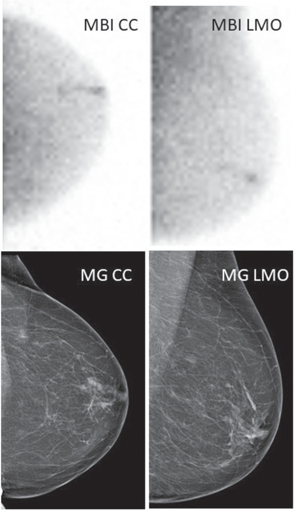
Results: Of the 96 cases of PND included, 78 were benign, and 18 (20%) corresponded to breast cancer (BC). Although CD and MBI were concordant in the BIRADS classification in 81% (78/96), half of BC were detected by MBI only. BC was located directly behind the nipple in a minority of patients (11%), meaning that MBI could significantly prevent futile central ductal excision. MBI presented higher sensitivity (83% vs. 33%) and negative predictive value (96% vs. 86%) than CD alone, with similar specificity (89% vs. 92%) and positive predictive value (63% vs. 50%). The area under the curve of MBI and CD was 0.86 ( P -value<0.001 [95% CI: 0.75-0.97]) and 0.63 ( P -value=0.091 [95% CI: 0.47-0.79]), respectively.
Conclusions: MBI showed good diagnostic accuracy for detecting BC in patients with PND with negative/indeterminate findings on CD imaging.
Keywords: Tc-99m sestamibi; breast cancer; breast imaging; gamma-camara scintigraphy; molecular breast imaging; nipple discharge.
Copyright © 2025 The Author(s). Published by Wolters Kluwer Health, Inc.
Conflict of interest statement
Conflicts of interest and sources of funding: none declared.
Figures


References
-
- Koskela A, Berg M, Pietiläinen T, et al. . Breast lesions causing nipple discharge: preoperative galactography-aided stereotactic wire localization. AJR Am J Roentgenol. 2005;184:1795–1798. - PubMed
-
- Goksel HA, Yagmurdur MC, Demirhan B, et al. . Management strategies for patients with nipple discharge. Langenbecks Arch Surg. 2005;390:52–58. - PubMed
-
- Newman HF, Klein M, Northrup JD, et al. . Nipple discharge. Frequency and pathogenesis in an ambulatory population. N Y State J Med. 1983;83:928–933. - PubMed
-
- Falkenberry SS. Nipple discharge. Obstet Gynecol Clin North Am. 2002;29:21–29. - PubMed
MeSH terms
Substances
LinkOut - more resources
Full Text Sources
Medical
