Review
. 2025 Apr 29;482(9):433-449.
doi: 10.1042/BCJ20253061.

Cytokines from parasites: manipulating host responses by molecular mimicry

Affiliations
Review

Cytokines from parasites: manipulating host responses by molecular mimicry

Rick M Maizels et al. Biochem J. .

Abstract

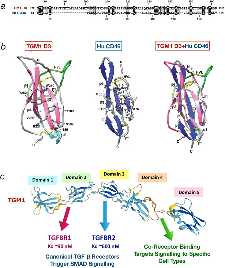
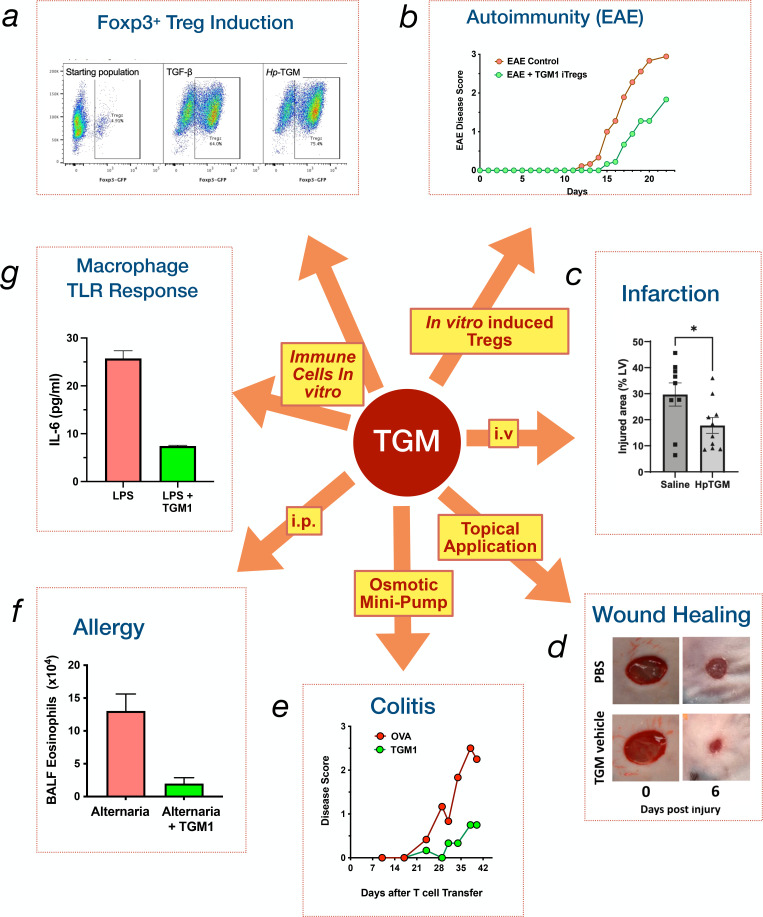
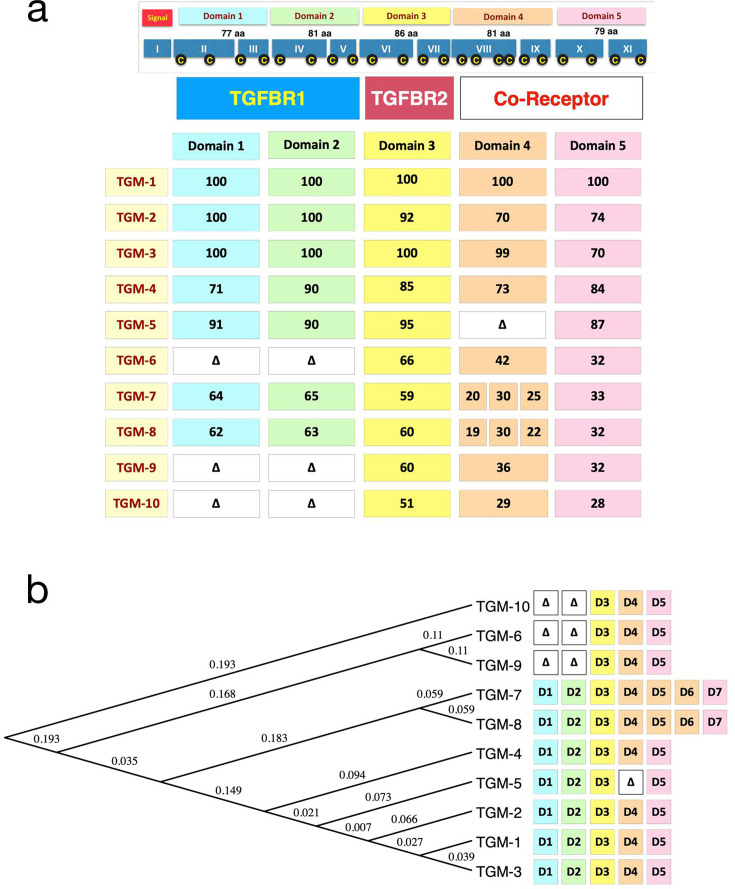
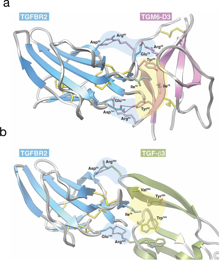
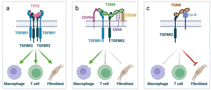
Helminth parasites have evolved sophisticated methods for manipulating the host immune response to ensure long-term survival in their chosen niche, for example, by secreting products that interfere with the host cytokine network. Studies on the secretions of Heligmosomoides polygyrus have identified a family of transforming growth factor-β (TGF-β) mimics (TGMs), which bear no primary amino acid sequence similarity to mammalian TGF-β, but functionally replicate or antagonise TGF-β effects in restricted cell types. The prototypic member, TGM1, induces in vitro differentiation of Foxp3+ T regulatory cells and attenuates airway allergic and intestinal inflammation in animal models. TGM1 is one of a family of ten TGM proteins expressed by H. polygyrus. It is a five-domain modular protein in which domains 1-2 bind TGFBR1, and domain 3 binds TGFBR2; domains 4-5 increase its potency by binding a co-receptor, CD44, highly expressed on immune cells. Domains 4-5 are more diverse in other TGMs, which bind co-receptors on cells such as fibroblasts. One variant, TGM6, lacks domains 1-2 and hence cannot transduce a signal but binds TGFBR2 through domain 3 and a co-receptor expressed on fibroblasts through domains 4-5 and blocks TGF-β signalling in fibroblasts and epithelial cells; T cells do not express the co-receptor and are not inhibited by TGM6. Hence, different family members have evolved to act as agonists or antagonists on various cell types. TGMs, which function by molecularly mimicking binding of the host cytokine to the host TGF-β receptors, are examples of highly evolved immunomodulators from parasites, including those that block interleukin (IL)-13 and IL-33 signalling, modulate macrophage and dendritic cell responses and modify host cell metabolism. The emerging panoply and potency of helminth evasion molecules illustrates the range of strategies in play to maintain long-term infections in the mammalian host.

Keywords: CD44; cytokines; evolution; immunomodulation; mimicry.


Conflict of interest statement

The authors declare that there are no competing interests associated with the manuscript.

Figures

Figure 1
Figure 1. Sequence and structural characteristics of TGM1 and CCP proteins.
(a) Alignment of TGM1 D3 with domain 2 of human CD46 [36]. Identical residues are shown in inverse shading, similar residues in shaded boxes. Amino acid positions in the respective proteins are indicated above and below the alignment. (b) Structural comparison of TGM1 D3 [36], left, with domain 2 of human CD46 (PDB 1CKL), centre and overlay of both structures, right. N- and C-termini of the domains are indicated, and positions of key amino acids. The four β sheets are indicated, as is the α1 helis. HVL, Hyper-Variable Loop. This part of the figure is reproduced from Ref. in the Journal of Biological Chemistry, in accordance with the republishing policy of the American Society for Biochemistry and Molecular Biology. (c) Alpha fold model of TGM1, indicating receptor specificities of the different domains, and the experimentally determined dissociation constants (Kd) for TGFBR1 and TGFBR2. Affinities of TGM4 and TGM6 for these receptors are given in Table 1.
Figure 2
Figure 2. Immunological functions of TGM1 in vitro and in vivo.
Schematic representing the various immune functions of TGM1. Clockwise from top left. (a) Induction of Foxp3 expression in murine [34,39 ] and human [40] T cells, representing the regulatory T cell (Treg) phenotype. (b) In vivo suppression of experimental autoimmune encephalomyelitis by adoptively transferred Tregs generated by in vitro treatment with TGM1, compared with TGF-β-induced Tregs [39]. (c) Reduction in cardiac injury during myocardial infarction by intravenous (i.v.) administration of TGM1 [39]. (d) Acceleration of skin wound healing in mice by topical application of TGM1 [41]. (e) Amelioration of colitis in RAG-deficient mice receiving Foxp3-negative T cells, in animals given subcutaneous osmotic minipumps releasing TGM1 [42]. (f) Suppression of allergic airway eosinophilic inflammation in bronchoalveolar lavage fluid (BALF) in mice receiving intranasal Alternaria alternata fungal allergen, and treated with intraperitoneal (i.p.) TGM1 [43]. (g) Inhibition of LPS-driven IL-6 release from murine bone marrow-derived macrophages in the presence of TGM1 [44]. TGF, transforming growth factor; TGM, transforming growth factor-β mimics.
Figure 3
Figure 3. TGM family member domain organisation and phylogenetic relationship.
(a) (Top section) Domain organisation of TGM1 and corresponding 11 exons in the H. polygyrus genome (Roman numerals); yellow C letters in black circles denote Cys residues. All disulphide bonds are intra-domain. (Main section) Organisation of domains in 10 family members, with percent amino acid identity of each domain to the corresponding domain of TGM1. Δ denotes absence of the domain(s). Note TGM7 and TGM8 have two additional domains, percentage identities shown are as indicated by colouring, with those with D4 of TGM1. Schematic is updated from Smyth et al [48] with corrected percent identities for D4-5 of TGM2 and the domains of TGM10 realigned. (b) Phylogenetic tree of the 10 full-length sequences of TGM family members. Numerals represent P values for branching topography. NB: For clarity, the TGM proteins are shown hyphenated (TGM-1), although normally the hyphen is omitted. TGF, transforming growth factor; TGM, transforming growth factor-β mimics.
Figure 4
Figure 4. Mimicry of TGFBR2 binding by TGM domain 3.
(a) Structure of TGM6-D3 (magenta) bound to the human TGFBR2 ectodomain (blue). Structure was determined using X-ray crystallography and was determined to a resolution of 1.4 Å [50]. Central hydrophobic interaction in which TGFBR2 Ile76 inserts into a pocket formed by TGM6-D3 Ile78, Tyr80 and Tyr93 is highlighted by yellow shading. Peripheral hydrogen-bonded ion pairs, TGFBR2 Asp55 and Glu78 with TGM6-D3 Arg95 and Arg38, respectively, and TGFBR2 Asp141 and Glu142 with TGM6-D3 Tyr80 and Arg82, respectively, that further stabilise the interaction and add specificity are highlighted by blue shading. (b) Structure of TGF-β3 (olive) bound to the human TGFBR2 ectodomain (blue). Structure was determined using X-ray crystallography and was determined to a resolution of 3.0 Å [53]. Central hydrophobic interaction in which TGFBR2 Ile76 inserts into a pocket formed by TGF-β3 Trp332, Tyr390 and Val392 is highlighted in yellow. Peripheral hydrogen-bonded ion pairs, TGFBR2 Asp55 with TGF-β3 Arg394 and TGFBR2 Glu142 with TGF-β3 Arg325, that further stabilise the interaction and add specificity are highlighted in yellow. TGF, transforming growth factor; TGM, transforming growth factor-β mimics.
Figure 5
Figure 5. Model of cell specific actions of TGM proteins.
Conceptual model comparing receptor interactions and cell specificity of TGF-β, TGM4 and TGM6. Created in BioRender. McSorley, HJ. (2025) https://BioRender.com/a55n320 (a) TGF-β acts as a homodimer to assemble a heterotetrameric complex of TGFBR12:TGFBR22 that signal to all cells expressing these receptors. (b) TGM4 ligates co-receptors including CD44, CD49d and CD206, while directly binding TGFBR1 and TGFBR2. D4-5 of TGM4 bind to CD44 and CD206; the domain binding to CD49d has not been conclusively identified. As a result of co-receptor interactions, TGM4 is selectively active on immune cells, particularly macrophages and does not activate fibroblasts with low or no expression of these co-receptors. (c) TGM6 lacks D1-2 and so cannot bind TGFBR1; it does however bind TGFBR2 together with a co-receptor (Co-R) that is different from those identified binding to TGM1 or TGM4. When added to cells expressing the appropriate co-receptor, such as fibroblasts, TGM6 acts as an antagonist of TGF-β signalling. TGF, transforming growth factor; TGM, transforming growth factor-β mimics.

Similar articles

Cited by

References

    1. Finlay B.B., McFadden G. Anti-immunology: evasion of the host immune system by bacterial and viral pathogens. Cell. 2006;124:767–782. doi: 10.1016/j.cell.2006.01.034. - DOI - PubMed
    1. Schmid-Hempel P. Parasite immune evasion: a momentous molecular war. trends ecol evol. 2008;23:318–326. doi: 10.1016/j.tree.2008.02.011. - DOI - PubMed
    1. Maizels R.M., Yazdanbakhsh M. Regulation of the immune response by helminth parasites: cellular and molecular mechanisms. Nat. Rev. Immunol. 2003;3:733–744. doi: 10.1038/nri1183. - DOI - PubMed
    1. McSorley H.J., Smyth D.J. IL-33 : A central cytokine in helminth infections. Semin. Immunol. 2021;53:101532. doi: 10.1016/j.smim.2021.101532. - DOI - PubMed
    1. Mayer D.A., Fried B. The role of helminth infections in carcinogenesis. Adv. Parasitol. 2007;65:239–296. doi: 10.1016/S0065-308X(07)65004-0. - DOI - PubMed

Publication types

MeSH terms

LinkOut - more resources