. 2025 Apr 25:18:5611-5625.
doi: 10.2147/JIR.S513823. eCollection 2025.

Taxus chinensis-Derived Nanovesicles Alleviate Mouse Colitis by Inhibiting Inflammatory Cytokines and Restoring Gut Microbiota

Affiliations

Taxus chinensis-Derived Nanovesicles Alleviate Mouse Colitis by Inhibiting Inflammatory Cytokines and Restoring Gut Microbiota

Qianyuan Gong et al. J Inflamm Res. .

Abstract

Background: Recent research has increasingly focused on plant-derived products as potential alternatives to chemotherapeutic agents, aiming to reduce side effects. Among these, plant-derived nanovesicles (NVs) have garnered significant attention for their potential in treating colitis.

Methods: In this study, we extracted NVs from the leaves (LNVs) and stems (SNVs) of Taxus, a well-known natural anti-cancer plant. The targeting ability of these NVs was evaluated in the mouse colon using an IVIS imaging system. Additionally, we assessed the therapeutic effects of these plant-derived NVs on ulcerative colitis in a mouse model.

Results: Our findings reveal that the NVs exhibit an ideal vesicle size of 150.0 nm and contain a rich array of lipids, functional proteins, and bioactive small molecules. In vitro anti-inflammatory experiments demonstrated that both LNVs and SNVs enhanced cell viability and reduced levels of pro-inflammatory cytokines. Importantly, neither LNVs nor SNVs induced significant cytotoxicity. In vivo, oral administration of LNVs and SNVs ameliorated colitis-related symptoms in mice and accelerated colitis resolution by suppressing the TLR4/MyD88/NF-κB pathway and reducing levels of pro-inflammatory cytokines, including IL-1β, IL-6, and TNF-α. Furthermore, 16S rDNA sequencing data suggested that LNVs play a crucial role in regulating gut microbiota.

Conclusion: Collectively, our findings suggest that plant-derived NVs from Taxus represent a promising novel natural nanomedicine for use as an anti-inflammatory agent in the treatment of colonic diseases.

Keywords: colitis; gut microbiota; plant-derived nanovesicles.


Conflict of interest statement

The authors declare no conflicts of interest in this work.

Figures

Figure 1
Figure 1
Characterization of TcNVs. (A) TcNVs were examined by transmission electron microscopy. Scale bar = 100 nm. Magnification: 120000X. (B) The particle size of TcNVs was analyzed by nanoparticle tracking analysis. (C) The lipid profiles of TcNVs under positive and negative mode. The lipid composition of TcNVs was determined by using a triple quadrupole mass spectrometer. The data are as percent ages of total signal for the molecular species determined after normalization of the signals in internal standards of the same lipid class. (D) The go secondary classification statistical charts of TcNVs.
Figure 2
Figure 2
In vivo distribution of DiR-labeled TcNVs. (A) Whole body imaging of the mice (healthy, LNVs treated and SNVs treated) at 2 h after oral administration of DiR-labeled TcNVs (1 mg/kg) by IVIS Spectrum Series imaging system. (B) The distribution of DiR-labeled TcNVs in the gastrointestinal tract captured by the IVIS system at 2 h after gavage. (C) The distribution of DiR-labeled TcNVs in the distal colons captured by the IVIS system at 2 h after gavage. (D) The relative fluorescence intensity in (C) was quantified using Image J software. The lowercase letters above bars indicated significant differences by one-way ANOVA. Data are shown as the mean ± SD (n=3). **p < 0.01 compared to the control group.
Figure 3
Figure 3
In vitro anti-inflammatory properties of TcNVs. (A and B) The LNVs (A) and SNVs (B) dose-dependently enhanced the cell vitality of LPS stimulated macrophages. Different amounts of TcNVs (1 µg/5 µg /10 µg /20 µg/mL, represented by SNV/LNV-1/5/10/20) were added to the THP-1 macrophages for 24 h. The cell vitality was tested by Cell Counting Kit-8. (CF) Result of inflammation-related cytokine IL-6 (C), IL-8 (D), IL-1β (E), and TNF-α (F) mRNA expression in LPS stimulated macrophages. Data are shown as the mean ± SD (n=3). **p < 0.01 compared to the negative control group; ##p < 0.01 compared to the positive control group.
Figure 4
Figure 4
TcNVs ameliorated the progression of DSS-induced mice colitis. (A) Experimental design to test the effects of TcNVs on DSS-induced colitis in mice (n = 8/group). (B) Result of body weight on TcNVs treatment in DSS-induced colitis. (C) Disease activity index (DAI) scores in each group. (D) Representative images of colon length in each group. (E) Statistical analysis of colon length in (D). Data are shown as the mean ± SD (n=8). **p < 0.01 and *p < 0.05 compared to the negative control group; ##p < 0.01 and #p < 0.05 compared to the positive control group.
Figure 5
Figure 5
TcNVs treatment improved mucosal inflammation of colitis tissues. (A and B) Representative colon histological sections stained with hematoxylin and eosin (H&E). Scale bar = 200 µm. (CE) Result of IL-1β, IL-6, and TNF-α in serum. Data are shown as the mean ± SD (n=3). **p < 0.01 compared to the negative control group; ##p < 0.01 compared to the positive control group.
Figure 6
Figure 6
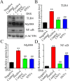
TcNVs suppresses the TLR4/MyD88/NF-κB signaling pathway. (A) Blots of TLR4, MyD88, p-NF-κB-p65 in the different experimental groups. (BD) Quantification the protein levels of TLR4 (B), MyD88 (C), p-NF-κB-p65 (D) in the different experimental treatment. Error bar represents SD (n = 3). **p < 0.01 compared to the negative control group; ##p < 0.01 compared to the positive control group.
Figure 7
Figure 7
Change of bacterial composition and community structure of gut microbiota in DSS-induced UC mice model under TcNVs interventions. (A) Venn diagram of all samples from four groups at ASV level. (B) Circos diagram of assigned main bacterial phyla between four groups. Top five phyla were shown. (C) Microbial compositions at genus level between groups. Top 15 genera were shown. (D) Principal coordinate analysis (PCoA) of Jaccard distance for DSS-induced UC mice model with different treatments (n = 6–7).
Figure 8
Figure 8
TcNVs treatments affected the taxonomic biomarkers and correlation with host in the DSS-induced UC mice. (A) Microbial biomarker analysis between the groups by Linear discriminant analysis effect size (LEfSe) analysis, LDA score (Log10) > 2.0. (B) Heatmap of Spearman correlation analysis between host data and top 30 genera that included the significantly enriched genera in TcNVs treatment groups. *FDR < 0.05. **FDR < 0.01. ***FDR < 0.001.

Similar articles

References

    1. Cantoro L, Monterubbianesi R, Falasco G, et al. The earlier you find, the better you treat: red flags for early diagnosis of inflammatory bowel disease. Diagnostics. 2023;13(20):3183. doi:10.3390/diagnostics13203183 - DOI - PMC - PubMed
    1. Upadhyay KG, Desai DC, Ashavaid TF, Dherai AJ. Microbiome and metabolome in inflammatory bowel disease. J Gastroenterol Hepatol. 2023;38(1):34–43. doi:10.1111/jgh.16043 - DOI - PubMed
    1. Zhang W, Michalowski CB, Beloqui A. Oral delivery of biologics in inflammatory bowel disease treatment. Front Bioeng Biotechnol. 2021;9:675194. doi:10.3389/fbioe.2021.675194 - DOI - PMC - PubMed
    1. Hanauer SB. New steroids for IBD: progress report. Gut. 2002;51:182–183. doi:10.1136/gut.51.2.182 - DOI - PMC - PubMed
    1. Zeng Z, He X, Li C, et al. Oral delivery of antioxidant enzymes for effective treatment of inflammatory disease. Biomaterials. 2021;271:120753. doi:10.1016/j.biomaterials.2021.120753 - DOI - PubMed

LinkOut - more resources