. 2024 Aug 12:2024:5785557.
doi: 10.1155/2024/5785557. eCollection 2024.

Genetic and Pathogenic Characteristics of an Emerging Highly Virulent Recombinant Lineage Korean Clade C PRRSV Strain

Affiliations

Genetic and Pathogenic Characteristics of an Emerging Highly Virulent Recombinant Lineage Korean Clade C PRRSV Strain

Jeongmin Suh et al. Transbound Emerg Dis. .

Abstract

A strain of porcine reproductive and respiratory syndrome virus (PRRSV) was isolated from lung tissue of a pig showing severe respiratory clinical signs from a farm in Gyeongsang province of South Korea. This PRRSV strain, designated as SNUVR220803, was classified within the lineage Korean clade C (LKC) based on a phylogenetic analysis of the ORF5 gene. A whole-genome analysis was conducted on the SNUVR220803 strain, which appears to be a recombinant between the PRRSV strains K07-2273 (part of LKC lineage) and Ingelvac MLV (part of Lineage 5). The Nsp2 amino acid sequence of this strain features a deletion of four additional amino acids, setting it apart from the typical Korean clades A, B, and C lineages. An animal inoculation experiment was conducted with 24 pigs divided into three groups: 12 pigs in the inoculated group, six in the sentinel group, and six in the negative control group. Inoculated pigs exhibited persisting hyperthermia (≥40.3°C) for 5 days, palpebral edema, and cyanosis. Subsequently, these pigs suffered from severe respiratory distress and cachexia, leading to a mortality rate of 58.3% (7 out of 12 pigs) at 14 days postinoculation (dpi). Body weight decreased post-SNUVR220803 strain infection in both the inoculated and sentinel groups. Gross pathology revealed noncollapsed lungs and serous effusion in the pericardial and peritoneal cavities. Microscopic analysis revealed severe interstitial pneumonia, while immunohistochemistry confirmed the presence of PRRSV antigen in the lungs, lymph nodes, thymus, kidneys, and the heart. Additionally, the levels of cytokines such as tumor necrosis factor-α (TNF-α), interferon-α (IFN-α), and IL-10 were significantly elevated in the plasma of infected pigs. These observations indicate that the LKC recombinant strain, combined with Lineage 5, possesses high virulence and infectivity as characterized by distinctive exudative lesions.


Conflict of interest statement

The author received support for the research presented in this article. However, this support did not exert any influence on the scientific content or stance of the research presented in this article.

Figures

Figure 1
Figure 1
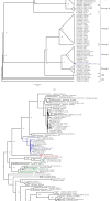
INDEL pattern of Nsp2 amino acid sequence of SNUVR220803. On the left side of the figure, the phylogenetic tree of Nsp2 amino acid sequences from 32 strains is shown, consisting of Korean PRRSV Type 2 strains and other representative strains from Lineages 1, 5, and 8. The major parental strain of the recombination event of the SNUVR220803 strain is shown with the green circle, and the blue circle represents the minor parental strain. Amino acid sequences from the right side show 300th to 800th amino acids in Nsp2 of each strain at the phylogenetic tree, which is a part of the HV region in the Nsp2 sequence. Deleted amino acids of the SNUVR220803 strain based on the VR-2332 strain are shown with red boxes, and the location of the deletion in the Nsp2 amino acid sequence is shown above the sequences with arrows and their numbers.
Figure 2
Figure 2
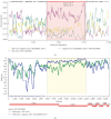
Phylogenetic trees based on ORF5 genes and whole genomes of the PRRSV-2 strains. In (a), 100 strains of PRRSV-2 are classified with their lineages in cartoon style based on ORF5 genes, and their location follows an increasing order. In (b), the phylogenetic tree is generated with whole genomes of the same strains as those of (a) and shown as a midpoint root tree. In terms of the recombination event, the major parental strain is marked with a green circle, and the minor parental strain is shown with a blue circle. Both trees are generated with 1,000 bootstrap tests, and the bootstrap numbers are noted at each of the diverging points of the phylogenetic trees.
Figure 3
Figure 3
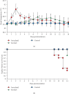
Recombination analysis of the SNUVR220803 strain between K07-2273 strain and Ingelvac MLV strain. (a) A recombination event ranging from 5,764 to 11,804 nt of SNUVR220803 based on the nucleotide sequence of VR-2332 is detected from the RDP4 software. Each color of the lines represents pairwise identity between the genomic sequence of SNUVR220803 and the parental strains. Expected recombination site is colored with a red box. (b) Similarity plot from the Simplot software is visualized, which represents the similarity between genomic sequences of the query strain (SNUVR220803) with the reference strains as the parental strains. The recombination site is also marked with a yellow color, and the arrows behind the similarity plot show the location of protein-coding sequences based on those of VR-2332.
Figure 4
Figure 4
Clinical scores: (a) respiratory clinical score, (b) activity score, and (c) body score. The significantly different groups (P < 0.05) are marked with different superscripts, including (A), (B), and (C).
Figure 5
Figure 5
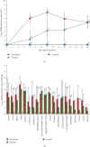
Rectal temperature and survival rate curve. (a) Rectal temperature data are shown, and groups significantly different (P < 0.05) are marked with alphabets, including (A), (B), and (C). (b) Kaplan–Meier survival rates.
Figure 6
Figure 6
Gross lesions found in pigs infected with SNUVR220803. Gross lesions of the pigs infected with SNUVR220803 are arranged. Each arrow of (a–f) indicates the following lesions found in live and necropsied pigs: (a) white nasal discharge, (b) eyelid edema, (c) and (d) cyanosis in extremities, (e) pericardium effusion, and (f) peritoneal effusion.
Figure 7
Figure 7
Quantification of PRRSV genomic copies in viremia and the tissue extracts. (a) The graph of logarithmic PRRSV genomic copy values in viremia of experimental pigs. Different superscripts including (A), (B), and (C) indicate statistically different groups (P < 0.05). (b) PRRSV genomic copies in 100 mg of each tissue samples.
Figure 8
Figure 8
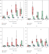
Cytokine levels in the plasma. The data of (a) TNF-α, (b) IFN- α, (c) IL-6, and (d) IL-10 are shown with box graphs. Data out of range are shown as dots separate from the box graphs. Different superscripts including (A), (B), and (C) indicate statistically different groups (P < 0.05).
Figure 9
Figure 9
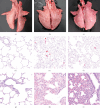
Lung lesions. (a–c) Show macroscopic lung lesions of the pigs in the control group and the inoculated group: control group (a), pigs in the inoculated group that deceased at 11 dpi (b), and at 14 dpi (c). (d–f) Show immunohistochemistry results in the lungs of the pigs in each experimental group: control group (d), pigs in the inoculated group that deceased at 11 dpi (e), and at 14 dpi (f). (g–i) Show HE staining of microscopic lung lesions of the pigs in each experimental group: control group (g), pigs in the inoculated group that deceased at 11 dpi (h), and at 14 dpi (i).
Figure 10
Figure 10
Immunohistochemistry about PRRSV antigen. The positive signals in immunohistochemistry of tissues from pigs infected with SNUVR220803 are arranged. The organs include (a) the lung, (b) the kidney, (c) lymph node, (d) thymus, (e) blood vessels in the cerebral sulcus, and (f) the heart. SR30 anti-PRRSV antibody is used for immunohistochemistry with alkaline phosphatase staining (red). The figures are in 200x magnification, and the insets are included (×400).

Similar articles

Cited by

References

    1. Cavanagh D. Nidovirales: a new order comprising Coronaviridae and Arteriviridae. Archives of Virology . 1997;142(3):629–633. - PubMed
    1. Walker P. J., Siddell S. G., Lefkowitz E. J., et al. Changes to virus taxonomy and the statutes ratified by the International Committee on Taxonomy of Viruses. Archives of Virology . 2020;165:2737–2748. doi: 10.1007/s00705-020-04752-x. - DOI - PubMed
    1. Shi M., Lam T. T.-Y., Hon C.-C., et al. Molecular epidemiology of PRRSV: a phylogenetic perspective. Virus Research . 2010;154(1-2):7–17. doi: 10.1016/j.virusres.2010.08.014. - DOI - PubMed
    1. Shi M., Lam T. T.-Y., Hon C.-C., et al. Phylogeny based evolutionary, demographical, and geographical dissection of North American type 2 porcine reproductive and respiratory syndrome viruses. Journal of Virology . 2010;84(17):8700–8711. doi: 10.1128/JVI.02551-09. - DOI - PMC - PubMed
    1. Kang H., Yu J. E., Shin J.-E., et al. Geographic distribution and molecular analysis of porcine reproductive and respiratory syndrome viruses circulating in swine farms in the Republic of Korea between 2013 and 2016. BMC Veterinary Research . 2018;14(1) doi: 10.1186/s12917-018-1480-6. - DOI - PMC - PubMed

MeSH terms

LinkOut - more resources