. 2024 Mar 22:2024:3009838.
doi: 10.1155/2024/3009838. eCollection 2024.

Galleria mellonella Invertebrate Model Mirrors the Pathogenic Potential of Mycoplasma alligatoris within the Natural Host

Affiliations

Galleria mellonella Invertebrate Model Mirrors the Pathogenic Potential of Mycoplasma alligatoris within the Natural Host

Alexandra M Burne et al. Transbound Emerg Dis. .

Abstract

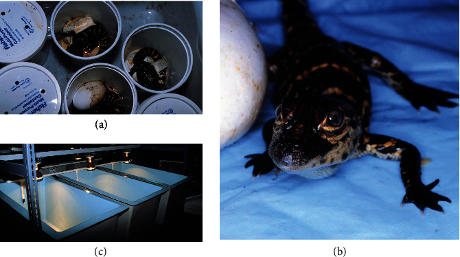
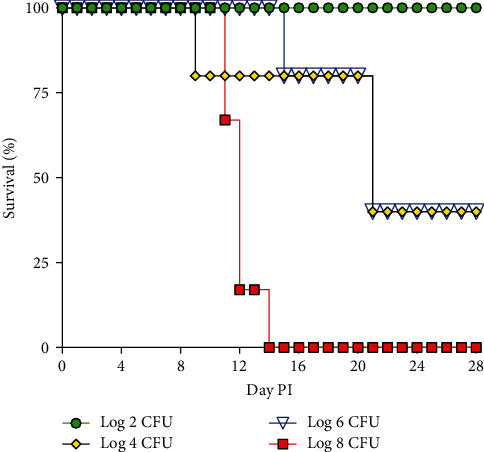
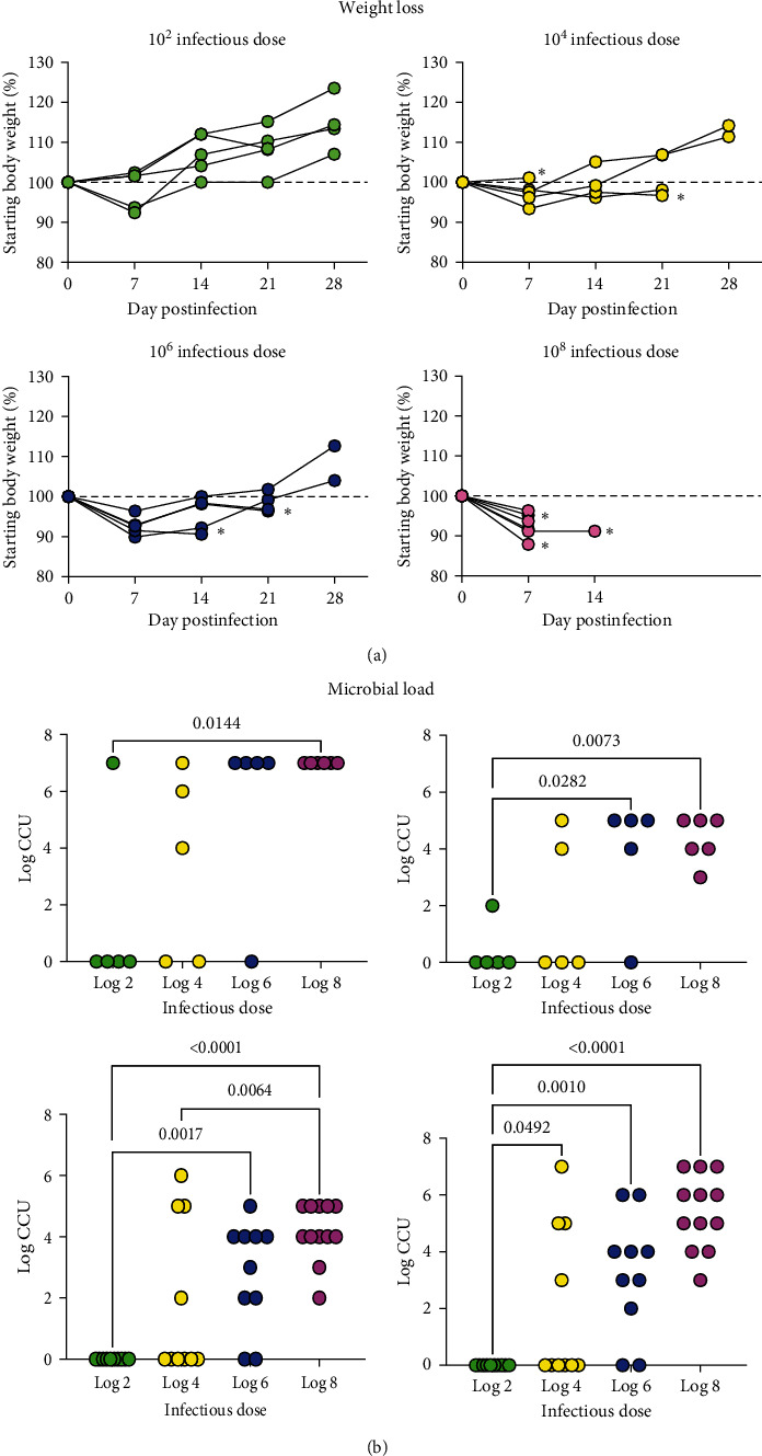
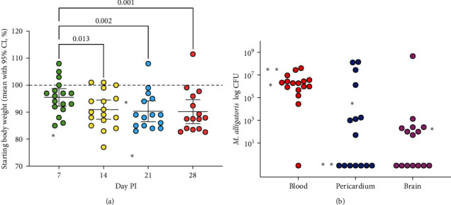
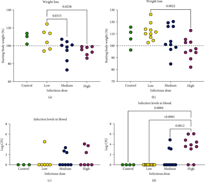
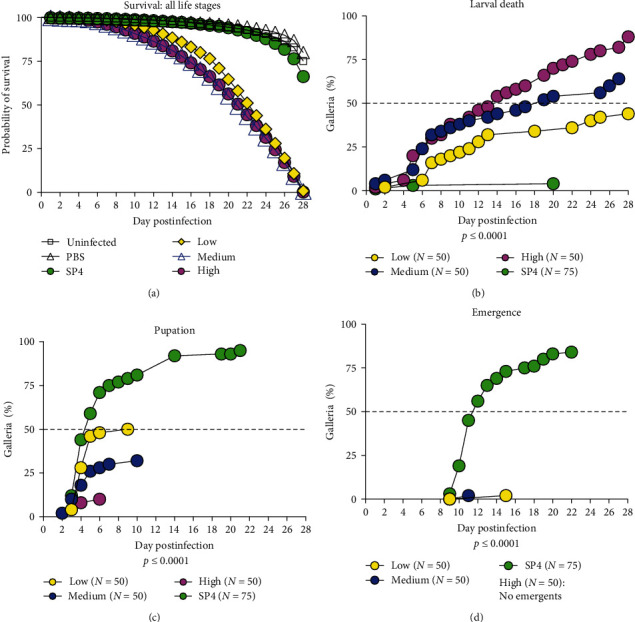
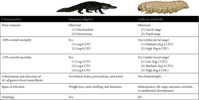
Most mycoplasmal infections result in chronic, clinically silent disease. In direct contrast, Mycoplasma alligatoris elicits a fulminant, multisystem disease in the natural host, Alligator mississippiensis (American alligator). The goals of the study were to better understand the disease in the natural host and to determine if the invertebrate model G. mellonella could serve as a surrogate alternate host. The survival of alligators infected intratracheally was dose dependent (p=0.0003), ranging from no mortality (102 CFU) to 100% mortality (108 CFU), with 60% mortality at the 104 and 105 CFU infectious dose. Microbial load in blood, joints, and brain was dose dependent, regardless of whether alligators were infected intratracheally or intravenously (p < 0.002). Weight loss was similarly impacted (p < 0.001). Experimental infection of the invertebrate Galleria mellonella mirrored the result in the natural host. In a dose response infection study, both larval survival curves and successful pupation curves were significantly different (p ≤ 0.0001) and dose dependent. Infected insects did not emerge as moths (p < 0.0001). Here, we describe the first study investigating G. mellonella as a surrogate model to assess the pathogenic potential of M. alligatoris. G. mellonella survival was dose dependent and impacted life stage outcome.


Conflict of interest statement

The authors declare that they have no conflicts of interest.

Figures

Figure 1
Figure 1
Husbandry of alligators. (a) Alligator eggs were incubated at 33°C. (b) Hatchlings were maintained in large plastic containers (c) tilted at an angle to provide approximately 7 cm water at the lower end and a dry area for basking at the higher end. Heat lamps provided 12-hr on/off cycles of light.
Figure 2
Figure 2
Survival curve for 22-week-old alligators inoculated intravenously with M. alligatoris. For infectious dose 102, 104, and 106 groups, n = 5; for 108 group, n = 6. No mortality was observed in controls (n = 5), data are not shown. Survival curves were analyzed by Kaplan–Meier and log-rank Mantel–Cox test for significance. Survival curves were significantly different, p=0.0003.
Figure 3
Figure 3
Weight loss (a) and bacterial colonization (b) in alligators infected intravenously with M. alligatoris. For infectious dose 102, 104, and 106 groups, n = 5; for 108 group, n = 6. Alligators were necropsied at time of euthanasia or 28 days postinfection. (a) The starting baseline weight is shown by the dashed line; all data are expressed in percentage of the starting body weight.   denotes last time point animal was weighed prior to euthanasia due to clinical criteria or mortality event. Note that in the 108 infectious dose, only one animal survived to the 14-day time point. (b) Culture results are shown for blood, brain, elbow, and knee. Both left and right joint sites are shown. All data are expressed as log CCU. Data were analyzed by one-way ANOVA; significant differences among groups were determined with Tukey's multiple comparison test. p values < 0.05 are shown. A linear trend was found for all sites (p  < 0.002).
Figure 4
Figure 4
Weight loss (a) and microbial load (b) in multiple body sites following intravenous (IV) infection with 106M. alligatoris. Three animals were euthanized at 6, 15, and 16D PI; all others were euthanized at D28 PI.   denotes the weight and microbial load of these three animals at time of euthanasia. (a) The starting baseline weight is shown by the dashed line; all data are expressed in percentage of the starting body weight. (b) Consistent bacteremia was found at necropsy with a subset of animals also infected in the brain. Note that two animals that were euthanized did not have a brain culture taken.
Figure 5
Figure 5
Weight loss (a, b) and bacterial colonization (c, d) in alligators infected intratracheally with M. alligatoris. Infectious doses used were low (102 and 103 CFU), medium (104 and 105 CFU), and high (106 and 107 CFU). Controls received sterile broth. Alligators were necropsied at Day 14 (control, n = 3; low, n = 6; medium, n = 8; high, n = 7) or Day 28 (control, n = 4; low, n = 10; medium, n = 10; high, n = 10) postinfection. Data were analyzed by one-way ANOVA; significant differences among groups were determined with Tukey's multiple comparison test. p values < 0.05 are shown. (a, b) The starting baseline weight is shown by the dashed line; all data are expressed in percentage of the starting body weight. A linear trend was found at both Day 14 (p=0.008) and Day 28 (p=0.002). Isolation of M. alligatoris from blood of intratracheally infected alligator is expressed as log CFU. At Day 28 PI (d), microbial load was significantly greater in the high infectious dose than all other groups, p  < 0.0001; there were no differences at Day 14 PI (c). A linear trend was found at Day 28 (p=0.0001) but not at Day 14 (p=0.205).
Figure 6
Figure 6
Representative gross and histological lesions in alligators infected with M. alligatoris. Lesions of the joints include (a) swollen footpads (arrows), (b) severe inflammation of the digit; the insert is a higher magnification of the area in hashed lines, showing infiltration of inflammatory cells and (c) inflammatory infiltrates into the joint space. Suppurative lesions of the coxofemoral joint (d) with synovitis (e) as well as occasional osteomyelitis (f). Gross fibrinous pericarditis (g) with inflammatory infiltrates (arrows) above the pericardium (h) and myometrium (i).
Figure 7
Figure 7
Impact of M. alligatoris on G. mellonella survival and transition across life stages. (a) Survival curves of control and M. alligatoris infected G. mellonella were significantly different (p ≤ 0.001, Kaplan–Meier and log-rank Mantel–Cox test). Differences in individual life stages were measured by percent larval mortality (b), pupation (c), or emergence (d). SP4-inoculated insects were the control group. (b) Larval mortality was significantly different (p ≤ 0.0001), one-way ANOVA. All groups were significantly different (Tukey's multiple comparisons), with the exception of larval mortality between the medium and high doses (p=0.1759). (c) Successful pupation among groups was significantly different (p ≤ 0.0001), Kruskal–Wallis test. Medium vs. SP4 (p=0.0074) and high vs. SP4 (p=0.0004) were significantly different, Dunn's multiple comparison test. (d) Emergence was significantly difference between SP4 controls and both low (p=0.0005) and medium (p=0.0028) infection doses, Dunn's multiple comparisons test. No insects in the high dose group were able to emerge.
Figure 8
Figure 8
Melanization of G. mellonella larva or pupae at time of death. Standard scoring for melanization: no melanization, score = 4; <3 black spots, score = 3; ≥3 dark spots, score = 2; fully melanized, score = 0. Melanization scores for mortality events are shown in the table and differed among groups, p=0.04, Chi-square. (a) Healthy larva; (b) healthy pupa; (c) larval–pupal intermediate; (d) larva; (e) larva; and (f) pupa.
Figure 9
Figure 9
Comparison of outcomes following experimental infection of the American alligator and G. mellonella. Overall mortality for alligators is defined as animals that died spontaneously or met euthanisa criteria. Overall mortality for Galleria is defined as death across all life stages; insect mortality was confirmed by the lack of movement after stimulation by gentle prodding with a sterile pipette tip.

Similar articles

References

    1. Arfi Y., Lartigue C., Sirand-Pugnet P., Blanchard A., Yount J. Beware of mycoplasma anti-immunoglobulin strategies. mBio . 2021;12(6) doi: 10.1128/mBio.01974-21.e0197421 - DOI - PMC - PubMed
    1. Askar H., Chen S., Hao H., et al. Immune evasion of Mycoplasma bovis. Pathogens . 2021;10(3) doi: 10.3390/pathogens10030297.297 - DOI - PMC - PubMed
    1. Dehon P. M., McGowin C. L. The immunopathogenesis of Mycoplasma genitalium infections in women: a narrative review. Sexually Transmitted Diseases . 2017;44(7):428–432. doi: 10.1097/OLQ.0000000000000621. - DOI - PMC - PubMed
    1. Dudek K., Nicholas R. A. J., Szacawa E., Bednarek D. Mycoplasma bovis infections-occurrence, diagnosis and control. Pathogens . 2020;9(8) doi: 10.3390/pathogens9080640.640 - DOI - PMC - PubMed
    1. Feberwee A., de Wit S., Dijkman R. Clinical expression, epidemiology, and monitoring of Mycoplasma gallisepticum and Mycoplasma synoviae: an update. Avian Patholology . 2022;51(1):2–18. doi: 10.1080/03079457.2021.1944605. - DOI - PubMed

MeSH terms

LinkOut - more resources