. 2024 Aug 24:2024:3680778.
doi: 10.1155/2024/3680778. eCollection 2024.

Development and Application of an RPA-Based Rapid Point-of-Care Testing (POCT) Method for the Detection of Feline Panleukopenia Virus

Affiliations

Development and Application of an RPA-Based Rapid Point-of-Care Testing (POCT) Method for the Detection of Feline Panleukopenia Virus

Liang Hong et al. Transbound Emerg Dis. .

Abstract

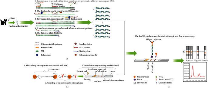
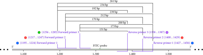
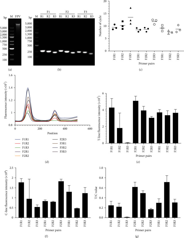
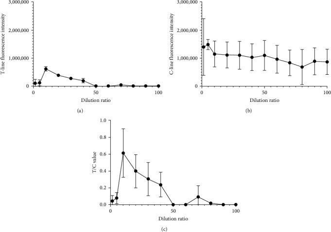
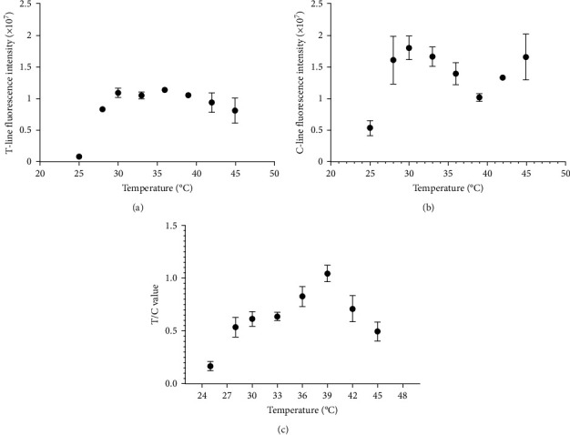
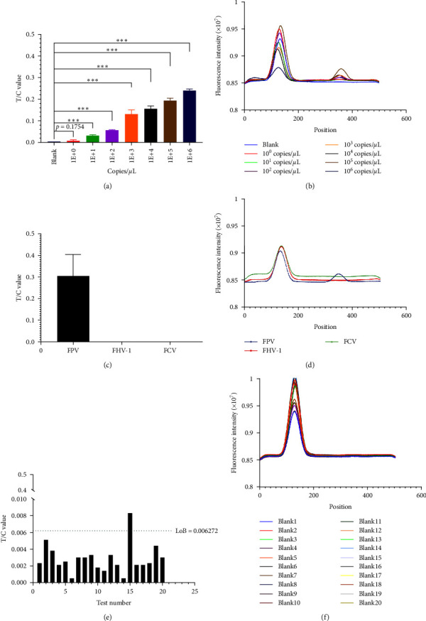
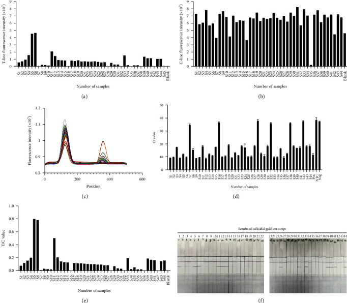
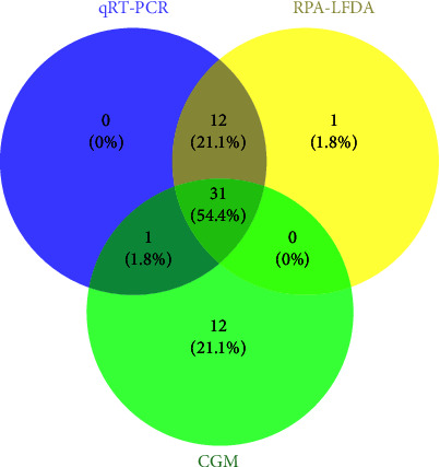
Feline panleukopenia (FP) is a highly prevalent and consequential disease that poses a substantial threat to both adult and juvenile cats across all geographical regions. The causative agent responsible for this disease is the feline panleukopenia virus (FPV). Therefore, it is imperative to develop a facile, efficient, and accurate detection method for FPV. Hence, a recombinase polymerase amplification-lateral flow dipstick assay (RPA-LFDA) method was specifically designed for the detection of FPV. The amplification process was optimized. This investigation focused on evaluating the expansion temperature detection system and revealed an optimal reaction temperature of 39°C. Then, primer combination screening involving nine groups identified F3R2 as the most effective primer set, while dilution ratio experiments determined that a 10-fold dilution yielded the best amplification products. Our findings demonstrated that the RPA-LFDA assay had an analytical sensitivity that was capable of detecting as low as 10 target copies per reaction. Furthermore, cross-reactivity tests demonstrated no interference between feline herpesvirus-1 (FHV-1) and feline calicivirus (FCV). To validate our newly developed method against existing techniques in clinical samples from three common sources on the market, we observed superior sensitivity and specificity compared to those of the colloidal gold method (CGM), with a higher positive detection rate using our nucleic acid detection system than CGM. Compared to qPCR as a reference standard, RPA-LFDA detected 39 out of 44 positive samples (including one false positive), whereas CGM detected 26 out of 44 positive samples. Based on the RPA-LFDA, the sensitivity was calculated to be 100%, the specificity was 83.33%, the mistake diagnostic rate was 16.67%, the omission diagnostic rate was 0%, and the overall accuracy reached 97.73%. Moreover, the positive coincidence rate was 97.44%, while the negative coincidence rate reached 100%. The agreement κ value was 0.8962. In conclusion, this approach exhibits greater sensitivity than CGM and offers greater convenience and cost-effectiveness than the qPCR methodology, making it a viable option for the clinical detection of FPV.


Conflict of interest statement

All authors affirm that they have no conflicts of interest or competing interests associated with this study.

Figures

Figure 1
Figure 1
An overview of the experimental approach employed by the RPA–LFDA system for the detection of feline panleukopenia virus (FPV). (a) Recombinase-based isothermal amplification was employed to amplify double-stranded DNA products labeled with fluorescein isothiocyanate (FITC) and biotin using DNA as the template. (b) Biomolecules were conjugated to microspheres via the coupling of biomolecules and subsequently applied onto the test strip, while the immobilization of streptavidin and goat anti-rabbit antibody occurred at the T-line and C-line positions simultaneously. (c) The RAPID product was applied to the strip, and the fluorescence intensity of the T and C-lines was quantified using a HIT-91A instrument.
Figure 2
Figure 2
Primer and probe design for the RPA–LFDA assay system. A set of three forward and three reverse primers was designed to target the FPV-VP2 gene, resulting in the amplification of nine nucleic acid fragments: F1R1 (261 bp), F1R2 (234 bp), F1R3 (192 bp), F2R1 (239 bp), F2R2 (212 bp), F2R3 (170 bp), F3R1 (200 bp), F3R2 (173 bp), and F3R3 (131 bp).
Figure 3
Figure 3
Results of FPV nucleic acid extraction and primer screening. (a) Results of FPV nucleic acid extraction by the TIANamp Virus DNA Kit. (b) Different primer combinations for PCR amplification. The amplified fragments were visualized by agarose gel electrophoresis. (c) Fluorescence intensity was monitored during amplification using a qRT-PCR instrument. SYBR Green I fluorescent dye was added to the RAPID amplification reaction mixture, and the resulting Ct value was subjected to statistical analysis. F3R2 had the smallest Ct value. (d) RPA amplification results under different primer combinations were tested, and the test strips were fully scanned by a HIT-91A instrument. The fluorescence intensities of the T-lines (e), C-lines (f) and T/C (g) were recorded and analyzed based on different primer combinations.
Figure 4
Figure 4
Dilution method for RPA product dilution. (a) Detection of the fluorescence intensity of the T-line was performed at various dilution ratios to determine the experimental outcomes. (b) Fluorescence intensity detection of the C-line was performed under various dilution ratios to obtain the experimental outcomes. (c) Fluorescence intensity detection of the T/C value was performed under various dilution ratios to obtain the experimental outcomes.
Figure 5
Figure 5
Optimal temperature screening for the RPA detection system. (a) Results of T-line fluorescence intensity under varying temperature conditions. (b) Results of C-line fluorescence intensity under varying temperature conditions. (c) Results of the T/C values under different temperature conditions.
Figure 6
Figure 6
Assessment of the analytical performance of the RPA–LFDA detection system. (a) Results of the T/C value for RPA–LFDA detection utilizing plasmids with varying copy numbers as templates ( p < 0.05). (b) The test strips were scanned after completion of the sensitivity testing process by a HIT-91A instrument. (c) The specificity of the RPA–LFDA system was assessed using FPV, FHV-1, and FCV nucleic acids. (d) The test strips were scanned after completion of the specificity testing process by a HIT-91A instrument. (e) To assess the detection limit of the RPA-LFDA detection system, a set of 20 blank samples was used as templates for isothermal amplification, followed by subsequent detection of the amplification products using the test strip. (f) The test strips were scanned after completion of the LoB testing process by a HIT-91A instrument. The symbol “ ∗∗∗” signifies the extremely significantly different p value = 0.0004.
Figure 7
Figure 7
The three assays were utilized to examine a total of 44 clinical samples. (a) The T values of the 44 specimens were ascertained through the application of the RPA–LFDA. (b) The C values of the 44 specimens were ascertained through the application of the RPA–LFDA. (c) The T/C values of the 44 specimens were ascertained through the application of the RPA–LFDA. (d) The test strip was subjected to scanning via the RPA–LFDA technique facilitated by the HIT-91A instrument. (e) Ct values were recorded using qRT-PCR of 44 samples for testing. (f) The results of 44 samples tested by the CGM assay were recorded.
Figure 8
Figure 8
Venn diagram of the number of FPVs identified via qRT-PCR, RPA–LFDA, and CGM.

Similar articles

References

    1. Roozitalab A., Elsakhawy O. K., Abouelkhair M. A., Matthijnssens J. M. Complete coding sequence of two feline panleukopenia virus strains isolated from domestic cats (Felis catus) in Tennessee, USA. Microbiology Resource Announcements . 2023;12(10) doi: 10.1128/MRA.00431-23.e0043123 - DOI - PMC - PubMed
    1. Pan S., Jiao R., Xu X., et al. Molecular characterization and genetic diversity of parvoviruses prevalent in cats in Central and Eastern China from 2018 to 2022. Frontiers in Veterinary Science . 2023;10 doi: 10.3389/fvets.2023.1218810.1218810 - DOI - PMC - PubMed
    1. Zhao S., Hu H., Lan J., et al. Characterization of a fatal feline panleukopenia virus derived from giant panda with broad cell tropism and zoonotic potential. Frontiers in Immunology . 2023;14 doi: 10.3389/fimmu.2023.1237630.1237630 - DOI - PMC - PubMed
    1. Kabir A., Habib T., Chouhan C. S., et al. Epidemiology and molecular characterization of feline panleukopenia virus from suspected domestic cats in selected Bangladesh regions. PLOS ONE . 2023;18(10) doi: 10.1371/journal.pone.0282559.e0282559 - DOI - PMC - PubMed
    1. Li S., Chen X., Hao Y., et al. Characterization of the VP2 and NS1 genes from canine parvovirus type 2 (CPV-2) and feline panleukopenia virus (FPV) in Northern China. Frontiers in Veterinary Science . 2022;9 doi: 10.3389/fvets.2022.934849.934849 - DOI - PMC - PubMed

MeSH terms

LinkOut - more resources