. 2025 Feb 3;10(4):1260-1273.
doi: 10.1016/j.ekir.2025.01.037. eCollection 2025 Apr.

Flow-Cytometric Quantification of Urine Kidney Epithelial Cells Specifically Reflects Tubular Damage in Acute Kidney Diseases

Affiliations

Flow-Cytometric Quantification of Urine Kidney Epithelial Cells Specifically Reflects Tubular Damage in Acute Kidney Diseases

Leonie Wagner et al. Kidney Int Rep. .

Abstract

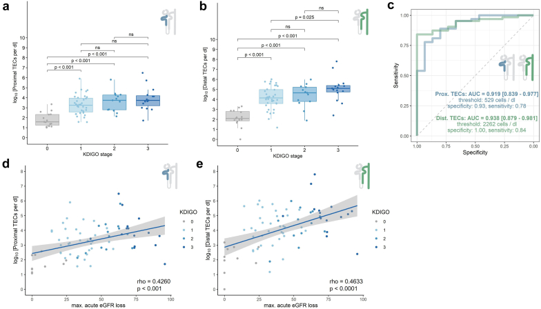
Introduction: Tubular injury is one of the main mechanisms driving acute kidney injury (AKI); however, clinicians still have a limited diagnostic repertoire to precisely monitor damage to tubular epithelial cells (TECs). In our previous study, we used single-cell sequencing to identify TEC subsets as the main components of the urine signature of AKI. This study aimed to establish TECs as clinical markers of tubular damage.

Methods: A total of 243 patients were analyzed. For sequencing, we collected 8 urine samples from patients with AKI and glomerular disease. We developed a protocol for the flow cytometric quantification of CD10/CD13+ proximal TECs (PTECs) and CD227/CD326+ distal TECs (DTECs) in urine by aligning urinary single-cell transcriptomes and TEC surface proteins using Cellular Indexing of Transcriptome and Epitope Sequencing (CITE-Seq). Marker combinations were confirmed in kidney biopsies. We validated our approach in 4 cohorts of 235 patients as follows: patients with AKI (n = 63), COVID-19 infection (n = 47), antineutrophil cytoplasmic autoantibody (ANCA)-associated vasculitis (AAV) with active disease or stable remission (n = 110), and healthy controls (n = 15).

Results: Our findings demonstrated that CD10/CD13 and CD227/CD326 adequately identified PTECs and DTECs, respectively. Distal urinary TEC counts correlate with the severity of AKI based on Kidney Disease: Improving Global Outcomes (KDIGO) stage and acute estimated glomerular filtration rate (GFR) loss in 2 separate cohorts and can successfully discriminate AKI from healthy controls and glomerular disease.

Conclusion: We propose that urinary CD227/CD326+ TEC count is a specific, noninvasive marker for tubular injury in AKI. Our protocol provides a basis for a deeper phenotypic analysis of urinary TECs.

Keywords: ANCA-associated vasculitis; acute kidney injury; flow cytometry; single-cell sequencing; tubular epithelial cells; urinalysis.


Figures

None
Graphical abstract
Figure 1
Figure 1
CITEseq in urinary single-cell sequencing shows loss of CD10 in urine. (a) Uniform manifold approximation and projection (UMAP) of 7575 single-cell RNA-sequencing urine cells from 7 individuals with AKI (n = 5), MGN (n = 1) and LN (n = 1). Cellular composition is individually diverse but can be broadly grouped into kidney cells (podocytes [PDCs] and distal and proximal tubular epithelial cells [DTECs or PTECs]), urogenital epithelial cells (UGECs), myeloid cells (MYEL), B and T lymphocytes (B/T LYMPHs) and erythrocytes (ERYs). (b–d) Quality control: Distribution of cells in UMAP by (b) genes per cell, (c) mitochondrial reads and (d) individual distribution. (e) Individual distribution of total transcriptomes and urinary cluster proportions. Irrespective of disease, predominantly DTECS and myeloid cells are found. (f) Dot plot of marker gene expression for each cell type. (g) RNA expression of CD10/MME, CD13/ANPEP, and CD326/EPCAM in tissue and urine and respective hashtag oligos in urine. CD326 is exclusively expressed on DTECs, CD13 on PTECs and myeloid cells, and CD10 is continuously expressed on podocytes. PTECs lose CD10 RNA expression in urine compared with tissue, while surface antigen markers stay positive. AKI, acute kidney injury; LN, lupus nephritis; MGN, membranous glomerular nephritis.
Figure 2
Figure 2
The amount of urinary TECs can be measured using flow cytometry. (a) Gating strategy for selecting cytokeratin positive single cells. Staining of CD10, CD13, CD326, and (b) CD227 compared with isotype-controls in postmortem kidney tissue, (c) postmortem ureteral tissue, and (d) urine. Note CD326 but not CD227 positivity in ureteral epithelial cells. (e,f) Fluorescence microscopic image of (e) purified CK+DAPI+CD13+CD10+ cells, and (f) purified CK+DAPI+CD326+CD227+ cells (blue = DAPI, green = cytokeratin). (g–j) Electron microscopic images of purified cytokeratin+CD227+ cells using a cell sorter. (g and detail h) Apoptotic cell with characteristic marginalization of condensed chromatin (yellow arrows in h) and intact plasma membrane. (i and j) Necrotic cells with chromatin clumping (red arrow in j) and loss of plasma membrane integrity as distinguishing feature from apoptosis (yellow arrow in j). TECs, tubular epithelial cells.
Figure 3
Figure 3
Flow cytometric quantification of proximal and distal TEC counts in patients with AKI. (a and b) (a) Proximal and (b) distal TEC counts shown for different KDIGO stages. (c) Receiver operator characteristic (ROC) curve for distinguishing AKI from healthy and inpatient controls. Area under the curve (AUC). Cut-offs for proximal and distal TEC counts were determined by Youden’s index. (d and e) Correlation of (d) proximal and (e) distal TECs and the maximal loss of eGFR during the course of AKI. AKI, acute kidney injury; eGFR, estimated glomerular filtration rate; TEC, tubular epithelial cell
Figure 4
Figure 4
Flow cytometric quantification of proximal and distal TEC counts in patients with COVID-19–AKI. (a and b) (a) Proximal and (b) distal TEC counts shown for different KDIGO stages and patients who develop AKI in the next 7 days. (c) Receiver operator characteristic (ROC) curve for distinguishing AKI from in-patient controls. Area under the curve (AUC). Cut-offs for proximal and distal TEC counts were determined by Youden’s index. (d and e) Correlation of (d) proximal (d) and (e) distal TECs and the maximal loss of eGFR during the course of AKI. AKI, acute kidney injury; dTEC, distal tubular epithelial cell; eGFR, estimated glomerular filtration rate; KDIGO, Kidney Disease: Improving Global Outcomes; pTEC, proximal tubular epithelial cell.
Figure 5
Figure 5
Flow cytometric quantification of proximal and distal TEC counts in patients with AAV. (a and c) (a) Proximal and (c) distal TEC counts in active and stable disease with and without renal involvement. (b and d) (b) Proximal and (d) distal TEC counts in active and stable disease in correlation with albuminuria. AAV, antineutrophil cytoplasmic autoantibody (ANCA)–associated vasculitis; AAV flare, active disease; AAV stable, inactive disease; TEC, tubular epithelial cells
Figure 6
Figure 6
Proximal and distal TECs show differences in renal damage patterns. (a and b) (a) Proximal and (b) distal TECs in healthy controls and different disease entities. (c) Receiver operator characteristic (ROC) curve for distinguishing AKI from healthy and inpatient controls and ANCA-associated vasculitis (AAV). Area under the Curve (AUC). Cut-offs for proximal and distal TEC counts were determined by Youden’s index. AKI, acute kidney injury; TEC, tubular epithelial cells.

References

    1. Hoste E.A.J., Kellum J.A., Selby N.M., et al. Global epidemiology and outcomes of acute kidney injury. Nat Rev Nephrol. 2018;14:607–625. doi: 10.1038/s41581-018-0052-0. - DOI - PubMed
    1. Cheng X., Wu B., Liu Y., Mao H., Xing C. Incidence and diagnosis of acute kidney injury in hospitalized adult patients: A retrospective observational study in a tertiary teaching Hospital in Southeast China. BMC Nephrol. 2017;18:203. doi: 10.1186/s12882-017-0622-6. - DOI - PMC - PubMed
    1. Wen Y., Yang C., Menez S.P., Rosenberg A.Z., Parikh C.R. A systematic review of clinical characteristics and histologic descriptions of acute tubular injury. Kidney Int Rep. 2020;5:1993–2001. doi: 10.1016/j.ekir.2020.08.026. - DOI - PMC - PubMed
    1. Hinze C., Kocks C., Leiz J., et al. Single-cell transcriptomics reveals common epithelial response patterns in human acute kidney injury. Genome Med. 2022;14:103. doi: 10.1186/s13073-022-01108-9. - DOI - PMC - PubMed
    1. Abdelhafez M., Nayfeh T., Atieh A., et al. Diagnostic performance of fractional excretion of sodium for the differential diagnosis of acute kidney injury: A systematic review and meta-analysis. Clin J Am Soc Nephrol. 2022;17:785–797. doi: 10.2215/CJN.14561121. - DOI - PMC - PubMed

LinkOut - more resources