

Comparison of commercial and in-house tissue-based and cell-based assays for the detection of autoantibodies targeting neuronal surface proteins: a prospective cohort study
- PMID: 40303411
- PMCID: PMC12038401
- DOI: 10.3389/fimmu.2025.1563877
Comparison of commercial and in-house tissue-based and cell-based assays for the detection of autoantibodies targeting neuronal surface proteins: a prospective cohort study
Abstract
Introduction: The detection of antibodies targeting neuronal antigens is a keystone for the diagnosis of autoimmune encephalitis (AE) and paraneoplastic neurological syndromes (PNS). This study aimed to compare the performance of a commercial tissue-based immunofluorescence assay (cIFA) to that of an inhouse IFA (hIFA) for the screening of autoantibodies targeting neuronal surface proteins in the cerebrospinal fluid (CSF) and to compare the performance of commercial cell-based assays (cCBA) to that of in-house CBA (hCBA) in serum samples.
Methods: Between March and June 2021, 2135 CSF samples and 524 serum samples from 2283 patients referred to the French Reference Center on PNS and AE were prospectively included. CSF samples were all tested using 3 different assays: cIFA, hIFA, and cCBA. Serum samples were all tested using at least 1 cCBA and 1 hCBA for the detection of the following autoantibodies: CASPR2, GABABR, and LGI1.
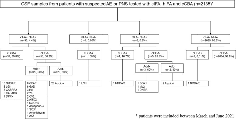
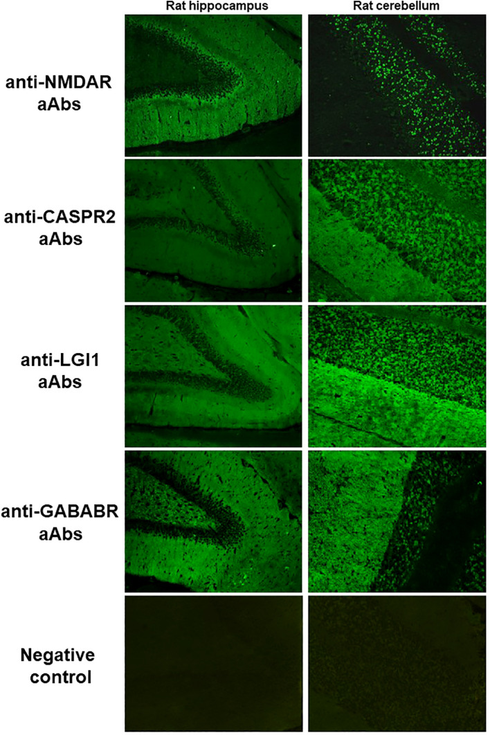
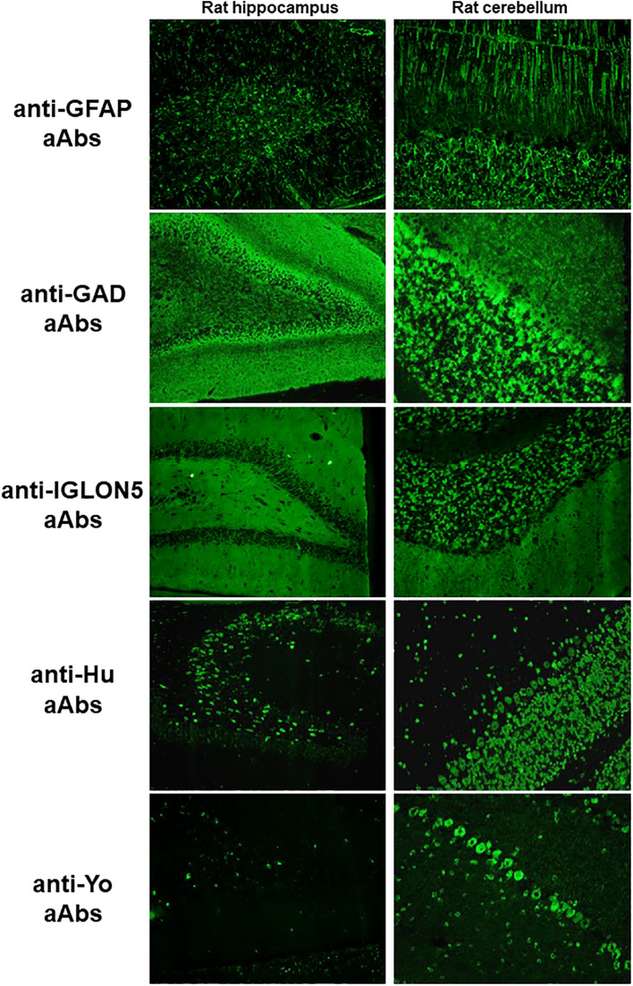
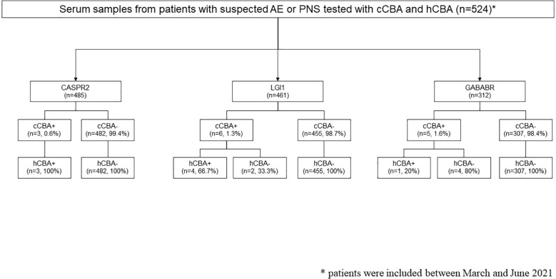
Results & discussion: Among the 2135 CSF tested, 93 (4.4%) were positive using both cIFA and hIFA, 1 (0.05%) was positive using only cIFA, and 6 (0.3%) were positive using only hIFA. Among the double-positive samples, 37 (39.8%) were positive using cCBA for the following autoantibodies: anti-NMDAR (n=16), -LGI1 (n=8), -CASPR2 (n=7), -GABABR (n=5), and -DPPX (n=1) autoantibodies. The remaining 56 (60.2%) double-positive samples were negative using cCBA and additional tests were performed to identify the autoantibodies according to the pattern observed on the IFA. The only sample positive using cIFA but negative using hIFA was positive for anti-LGI1 autoantibodies using cCBA. Among the 6 samples negative using cIFA but positive using hIFA, only one sample was positive with cCBA for anti-NMDAR autoantibodies. These data indicate that, in CSF, cIFA and hIFA performed similarly for the detection of autoantibodies targeting neuronal surface proteins.Regarding serum samples, cCBA and hCBA were both positive in 3 patients for CASPR2, 4 patients for LGI1, and 1 patient for GABABR. A positive cCBA and negative hCBA was observed in 2 patients for LGI1 and 4 patients for GABABR. A lack of specificity of GABABR cCBA is suspected as CSF explorations were negative in 3 of these patients and none presented clinical features highly suggestive of AE.
Keywords: autoantibodies; autoimmune encephalitis; cell-based assay; diagnostic test; immunofluorescence assays; paraneoplastic neurological syndromes; tissue-based assay.
Copyright © 2025 Goncalves, Benaiteau, Rogemond, Closs, Pinto, Dhairi, Villard, Picard, Fabien and Honnorat.
Conflict of interest statement
The authors declare that the research was conducted in the absence of any commercial or financial relationships that could be construed as a potential conflict of interest.
Figures
Similar articles
-
Limitations of a Commercial Assay as Diagnostic Test of Autoimmune Encephalitis.Front Immunol. 2021 Jun 29;12:691536. doi: 10.3389/fimmu.2021.691536. eCollection 2021. Front Immunol. 2021. PMID: 34267758 Free PMC article.
-
Assessing Commercial Tissue-Based Assays for Autoimmune Neurologic Disorders (II): Antibodies to Surface Antigens.Neurol Neuroimmunol Neuroinflamm. 2025 Jul;12(4):e200406. doi: 10.1212/NXI.0000000000200406. Epub 2025 May 20. Neurol Neuroimmunol Neuroinflamm. 2025. PMID: 40393020 Free PMC article.
-
Autoimmune Encephalitis and Paraneoplastic Neurologic Syndromes: A Nationwide Study on Epidemiology and Antibody Testing Performance.Neurol Neuroimmunol Neuroinflamm. 2024 Nov;11(6):e200318. doi: 10.1212/NXI.0000000000200318. Epub 2024 Oct 28. Neurol Neuroimmunol Neuroinflamm. 2024. PMID: 39467237 Free PMC article.
-
Diagnostic tools for immune causes of encephalitis.Clin Microbiol Infect. 2019 Apr;25(4):431-436. doi: 10.1016/j.cmi.2018.12.012. Epub 2018 Dec 22. Clin Microbiol Infect. 2019. PMID: 30583056 Review.
-
[Clinical Phenomenology of Autoimmune Encephalitis].Fortschr Neurol Psychiatr. 2016 May;84(5):271-80. doi: 10.1055/s-0042-104194. Epub 2016 Jun 14. Fortschr Neurol Psychiatr. 2016. PMID: 27299786 Review. German.
References
Publication types
MeSH terms
Substances
Supplementary concepts
LinkOut - more resources
Full Text Sources
Medical