. 2023 Apr 24:2023:9248581.
doi: 10.1155/2023/9248581. eCollection 2023.

A Bivalent Bacterium-like Particles-Based Vaccine Induced Potent Immune Responses against the Sudan Virus and Ebola Virus in Mice

Affiliations

A Bivalent Bacterium-like Particles-Based Vaccine Induced Potent Immune Responses against the Sudan Virus and Ebola Virus in Mice

Shengnan Xu et al. Transbound Emerg Dis. .

Abstract

Ebola virus disease (EVD) is an acute viral hemorrhagic fever disease causing thousands of deaths. The large Ebola outbreak in 2014-2016 posed significant threats to global public health, requiring the development of multiple medical measures for disease control. Sudan virus (SUDV) and Zaire virus (EBOV) are responsible for severe disease and occasional deadly outbreaks in West Africa and Middle Africa. This study shows that bivalent bacterium-like particles (BLPs)-based vaccine, SUDV-EBOV BLPs (S/ZBLP + 2 + P), generated by mixing SUDV-BLPs and EBOV-BLPs at a 1 : 1 ratio, is immunogenic in mice. The SUDV-EBOV BLPs induced potent immune responses against SUDV and EBOV and elicited both T-helper 1 (Th1) and T-helper 2 (Th2) immune responses. The results indicated that SUDV-EBOV BLPs-based vaccine has the potential to be a promising candidate against SUDV and EBOV infections and provide a strategy to develop universal vaccines for EVD.


Conflict of interest statement

The authors declare that they have no conflicts of interest.

Figures

Figure 1
Figure 1
Schematic view of EBOV eGP-PA with the GP ectodomain of EBOV, linker, and PA.
Figure 2
Figure 2
Identification of EBOV eGP-PA fusion Protein. The CPE in sf9 cells infected with rBV-EBOV eGP-PA (a) and uninfected sf9 cells (b) was observed. The PCR amplification of rBV-EBOV eGP-PA genome with oligonucleotide primers EBOV-eGP-F/Linker-PA-R (c); M: DNA marker; lane 1: control baculovirus; lane 2: the P3 recombinant baculoviruses. IFA detection of the expression of EBOV eGP-PA in baculovirus-infected Sf9 insect cells (magnification of microscopy images, 200x). The sf9 cells, fed in 24 well plates, were incubated with mouse anti-EBOV GP monoclonal antibody and then detected using conjugated goat antimouse IgG (d, e). WB analysis of EBOV eGP-PA fusion protein expression patterns (f). M: protein marker; lane 1: culture medium supernatant; lane 2: precipitate following supersonic schizoanalysis; lane 3: supernatant following supersonic schizoanalysis. GP: 100 kDa; PA: 30 kDa.
Figure 3
Figure 3
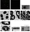
Identification of BLPs loaded with EBOV eGP-PA. IFA analysis of the BLPs loaded with EBOV eGP-PA (magnification of microscopy images, 1000x). BLPs loaded with EBOV eGP-PA (a); BLPs without fusion protein loaded (b). WB analysis of EBOV eGP-PA fusion protein binding with BLPs (c). M: protein marker; lane 1: the precipitate of fusion protein bound to BLPs; lane 2: the supernatant of fusion protein bound to BLPs; lane 3: BLPs. Transmission electron microscopy analysis of L. lactis without (d) or with TCA treated (e) and BLPs without fusion protein loaded (f) and BLPs loaded with EBOV eGP-PA fusion protein (g). WB analysis of the binding amount of the fusion protein on BLPs (h) SDS-PAGE analysis of the maximum binding capacity of EBOV eGP-PA fusion protein to BLPs (i). M: protein marker; 1: combined SUDV eGP-PA; 2: combined EBOV eGP-PA.
Figure 4
Figure 4
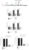
Serum antibody responses induced by SUDV-EBOV BLPs. Overall study design (a). Mice were intramuscularly immunized with S/ZBLP + 2 + P, SBLP + 2 + P, ZBLP + 2 + P, 2 + P, and PBS 3 weeks apart. Serum samples of immunized mice were collected by retro-orbital plexus puncture at 2, 4, and 5 weeks after prime immunization, respectively. To assess cell-mediated immune responses induced by SUDV-EBOV BLPs, spleens were collected 8 d after boost immunization. The horizontal dotted line in the figure indicates the limit of detection (LOD). Data are shown as the mean ± SD and were analyzed by one-way ANOVA. The SUDV-and EBOV-specific IgG antibody levels induced by SUDV-EBOV BLPs were analyzed using indirect ELISA (b, c). Neutralizing antibody levels of serum samples were determined using pseudovirion neutralization assay (d, e).
Figure 5
Figure 5
SUDV GP- and EBOV GP-specific IgG antibody subtypes responses induced by SUDV-EBOV BLPs. Serum samples were collected to detect SUDV GP- and EBOV GP-specific IgG1 and IgG2a antibody levels using indirect ELISA and IgG1, and IgG2a antibody responses were displayed as the end-point dilution titers. The ratios of IgG2a/IgG1 were calculated. Data are shown as the mean ± SD and were analyzed by one-way ANOVA. SUDV GP-IgG1 and IgG2a antibody levels (a) and IgG2a/IgG1 ratios (c) EBOV GP-specific IgG1 and IgG2a antibody levels (b) and IgG2a/IgG1 ratios (d)
Figure 6
Figure 6
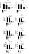
Cell-mediate responses induced by SUDV-EBOV BLPs vaccine. Data are shown as the mean ± SD and were analyzed by one-way ANOVA. The splenocytes, isolated from mice, were used to perform a splenocyte proliferation assay and detect the antigen-specific IFN-γ, IL-4, and TNF-α secretion levels using ELISpot. The stimulation index of the splenocytes was detected using CCK-8 solution by stimulating the splenocytes with purified SUDV GP (a) and EBOV GP (b), respectively. Splenocytes secreting IFN-γ, IL-4, and TNF-α were quantified using an ELISpot assay after SUDV GP (c–e) and EBOV GP (f–h) stimulation.
Figure 7
Figure 7
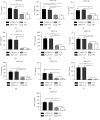
Cytokine secretion of splenocyte culture supernatant. Data are shown as the mean ± SD and were analyzed by one-way ANOVA. Cytokine secretion levels in the supernatant were measured with commercial ELISA kits. The concentration of IL-4, IL-10, IL-2, IFN-γ, and TNF-α (a–e) was measured after stimulation with SUDV GP and EBOV GP (f–j).

Similar articles

References

    1. Centers for Disease Control and Prevention. Ebola(Eboal virus disease) 1976. https://www.cdc.gov/vhf/ebola/history/distribution-map.html .
    1. Di Paola N., Sanchez-Lockhart M., Zeng X., Kuhn J. H., Palacios G. Viral genomics in Ebola virus research. Nature Reviews Microbiology . 2020;18(7):365–378. doi: 10.1038/s41579-020-0354-7. - DOI - PMC - PubMed
    1. Ghosh S., Saha A., Samanta S., Saha R. P. Genome structure and genetic diversity in the Ebola virus. Current Opinion in Pharmacology . 2021;60:83–90. doi: 10.1016/j.coph.2021.06.010. - DOI - PubMed
    1. Goldstein T., Anthony S. J., Gbakima A., et al. The discovery of Bombali virus adds further support for bats as hosts of ebolaviruses. Nat Microbiol . 2018;3(10):1084–1089. doi: 10.1038/s41564-018-0227-2. - DOI - PMC - PubMed
    1. Murin C. D., Gilchuk P., Crowe J. E., Ward A. B. Structural biology illuminates molecular determinants of broad ebolavirus neutralization by human antibodies for pan-ebolavirus therapeutic development. Frontiers in Immunology . 2021;12 doi: 10.3389/fimmu.2021.808047.808047 - DOI - PMC - PubMed

LinkOut - more resources