

Quantitative SISCOM assessment for epileptogenic zone localization: insights from a multicenter study comparing two software platforms in temporal lobe epilepsy
- PMID: 40303886
- PMCID: PMC12037379
- DOI: 10.3389/fneur.2025.1552774
Quantitative SISCOM assessment for epileptogenic zone localization: insights from a multicenter study comparing two software platforms in temporal lobe epilepsy
Abstract
Introduction: Pharmacoresistant epilepsy affects around one-third of individuals with epilepsy, requiring precise diagnosis, particularly in cases where surgical resection of the epileptogenic zone (EZ) is an option. Functional imaging techniques, such as ictal-interictal subtraction SPECT coregistered to MRI (SISCOM), have proven useful in pre-surgical evaluation by improving EZ localization accuracy. However, the widespread use of SISCOM is limited by the high costs and technical complexity of commercial software. Statistical Parametric Mapping (SPM) has been demonstrated to be a viable alternative for SISCOM analysis, displaying the potential for cost-effective EZ localization.
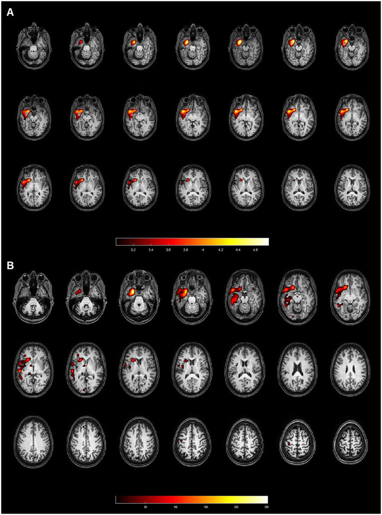
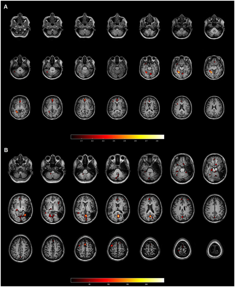
Materials and methods: In this retrospective study, we evaluated patients with pharmacoresistant temporal lobe epilepsy from two reference centers of epilepsy in Brazil, who underwent ictal and interictal SPECT imaging as part of their pre-surgical evaluation, achieving favorable outcomes (Engel I or II) after surgical resection. The EZ reference standard was determined according to anatomopathological findings and good clinical outcomes. SISCOM was performed using a semi-automated approach with Statistical Parametric Mapping (SPM) and a proprietary software - Analyze. Data from each method were compared to the EZ reference standard and classified as concordant, partially concordant, or discordant.
Results: We included 20 patients, 14 (70%) with left temporal lobe epilepsy and six (30%) with right temporal lobe epilepsy. Hippocampal sclerosis was the most common pathology (80%). Both SPM and Analyze were concordant with the EZ reference standard in 14 cases (70%), showing no difference in sensitivity between the methods. However, SPM generated smaller, more localized clusters, while Analyze produced larger clusters with broader spatial coverage. Concordance between the two methods was poor (Kappa = 0.0179), reflecting methodological differences.
Conclusion: This study evidences technical differences between SISCOM performed with SPM and Analyze, but with similar sensitivity (70%) for EZ localization. Further studies with larger sample sizes are required to confirm these findings. The data presented here suggest that SISCOM-SPM, due to its rapid and semi-automated workflow, may offer a practical and accessible alternative to proprietary software for epilepsy surgical planning.
Keywords: SISCOM; SPECT; epileptogenic zone; pharmacoresistant epilepsy; quantitative analysis; temporal lobe epilepsy.
Copyright © 2025 Oliveira Young, de Campos, de Souza, Brunetto, Gualberto, Alexandre-Santos, de Oliveira, Alvim, Cendes, Etchebehere, Wichert-Ana and Amorim.
Conflict of interest statement
The authors declare that the research was conducted in the absence of any commercial or financial relationships that could be construed as a potential conflict of interest.
Figures
Similar articles
-
Clinical Usefulness of SISCOM-SPM Compared to Visual Analysis to Locate the Epileptogenic Zone.Front Neurol. 2020 May 29;11:467. doi: 10.3389/fneur.2020.00467. eCollection 2020. Front Neurol. 2020. PMID: 32547479 Free PMC article.
-
The effect of injection time on rates of epileptogenic zone localization using SISCOM and STATISCOM.Epilepsy Behav. 2021 May;118:107945. doi: 10.1016/j.yebeh.2021.107945. Epub 2021 Apr 10. Epilepsy Behav. 2021. PMID: 33845344
-
Resecting critical nodes from an epileptogenic circuit in refractory focal-onset epilepsy patients using subtraction ictal SPECT coregistered to MRI.J Neurosurg. 2016 Dec;125(6):1565-1576. doi: 10.3171/2015.6.JNS141719. Epub 2016 Mar 18. J Neurosurg. 2016. PMID: 26991384
-
Neuronuclear assessment of patients with epilepsy.Semin Nucl Med. 2008 Jul;38(4):227-39. doi: 10.1053/j.semnuclmed.2008.02.004. Semin Nucl Med. 2008. PMID: 18514079 Review.
-
Use of Innovative SPECT Techniques in the Presurgical Evaluation of Patients with Nonlesional Extratemporal Drug-Resistant Epilepsy.Mol Imaging. 2021 Mar 2;2021:6614356. doi: 10.1155/2021/6614356. eCollection 2021. Mol Imaging. 2021. PMID: 33746629 Free PMC article. Review.
References
-
- Devous MD, Sr, Thisted RA, Morgan GF, Leroy RF, Rowe CC. SPECT brain imaging in epilepsy: a meta-analysis. J Nucl Med. (1998) 39:285–93. PMID: - PubMed
LinkOut - more resources
Full Text Sources