. 2025 Feb 13:48:43-54.
doi: 10.1016/j.bioactmat.2025.02.004. eCollection 2025 Jun.

MiR-19-loaded oxidative stress-relief microgels with immunomodulatory and regeneration functions to reduce cardiac remodeling after myocardial infarction

Affiliations

MiR-19-loaded oxidative stress-relief microgels with immunomodulatory and regeneration functions to reduce cardiac remodeling after myocardial infarction

Kai Wang et al. Bioact Mater. .

Abstract

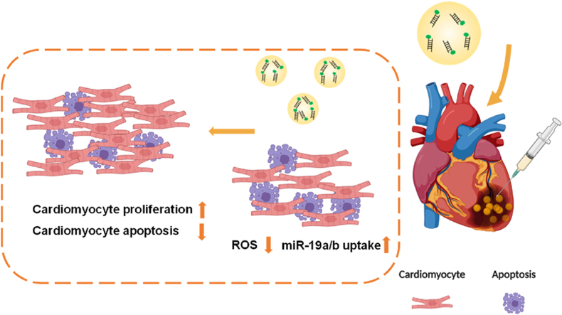
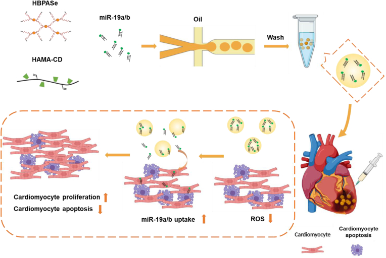
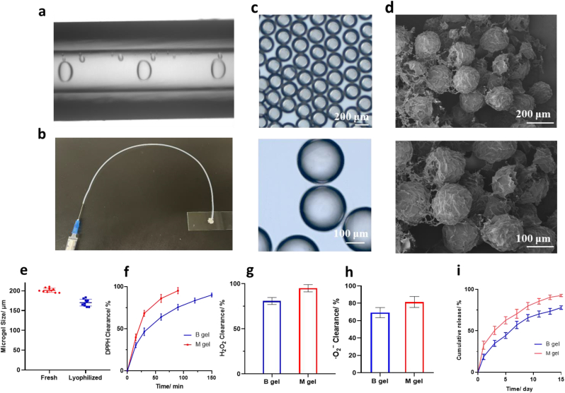
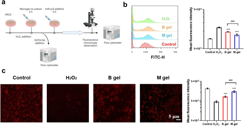
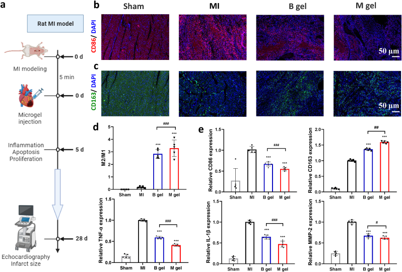
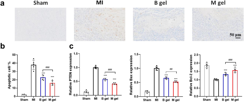
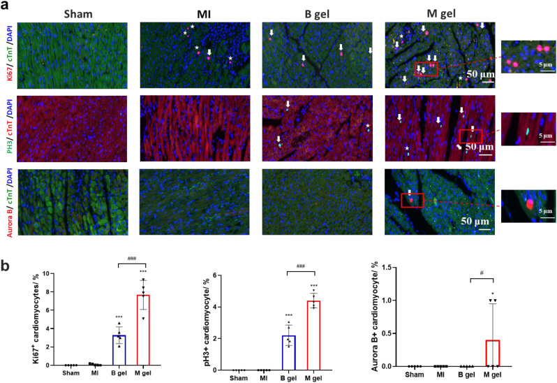
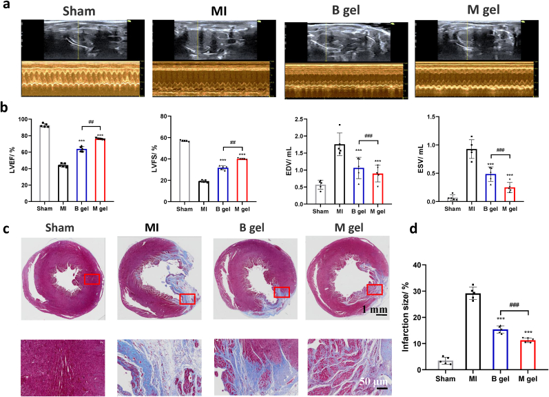
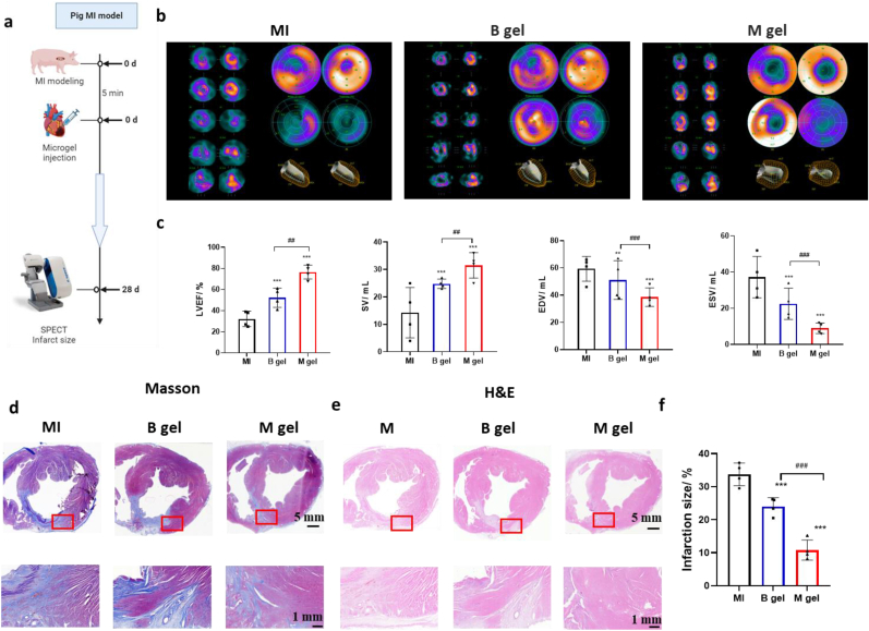
Regeneration therapeutic strategy by microRNAs for boosting cardiomyocyte proliferation in treating myocardial infarction (MI) has the challenges of efficient delivery, and toxicity and risk of sudden death. Herein, oxidative stress-relief microgels were developed for miR-19a/b delivery, modulation of inflammatory tissue microenvironment, promotion of cardiomyocyte proliferation, and maintenance of heart function post MI. The cholesterol-modified miR-19a/b was encapsulated into the cavity of β-cyclodextrin in selenoketal-containing microgels. The microgels could effectively scavenge typical reactive oxygen species (ROS), and down-regulate the intracellular ROS level and the levels of typical inflammatory factors. The microgels could improve the acute inflammatory microenvironment for better cardiomyocyte survival and cellular uptake of miR-19a/b, leading to significant promotion of cardiomyocyte proliferation in vivo. In the rat and minipig models of MI, the microgels most effectively inhibited the acute inflammatory response and reduced the cardiomyocytes apoptosis, resulting in a significant improvement of cardiac function and restriction of pathological remodeling post MI, and thereby best heart function revealed by echocardiography and histological analysis.

Keywords: Cardiomyocyte regeneration; Inflammation; Microgels; Myocardial infarction; microRNA.


Conflict of interest statement

The authors declare that they have no known competing financial interests or personal relationships that could have appeared to influence the work reported in this paper.

Figures

Image 1
Graphical abstract
Scheme 1
Scheme 1
Schematic illustration of ROS-responsive microgels loaded with miR-19a/b (M gel) for promoting cardiomyocyte proliferation in vivo. Preparation of M gel building blocks by encapsulating miR-19a/b into ROS-scavenging microgels in a microfluidic device. The microgels are formed by crosslinking the precursor solutions of HBPASe and HAMA-CD macromolecules. The miR-19a/b modified with cholesterol is loaded into the cavity of cyclodextrin of HAMA-CD. This system can sensitively scavenge the locally overproduced ROS in the MI microenvironment, continuously release the miR-19a/b, and improve cellular uptake of miR-19a/b for efficiently stimulating the proliferation of cardiomyocytes and reducing apoptosis.
Fig. 1
Fig. 1
Physiochemical properties of ROS-responsive microgels loaded with miR-19a/b. (a) Homogeneous droplets formed at a flow focusing junction of the microfluidic channel. (b) Injection of the M gel through a catheter. (c) Optical microscopy images of freshly prepared microgels. (d) SEM images of freeze-dried microgels. (e) Size of freshly prepared and lyophilized microgels. (f) Clearance of DPPH radicals, (g) H2O2 and (h) ·O2 by the bulk hydrogel (B gel) and M gel. (i) Release of cholesterol-modified miR-19a/b from the B gel and M gel in 100 mM H2O2 containing DMEM. n = 3 per group.
Fig. 2
Fig. 2
M gel protects cells against oxidative stress and maintains cellular uptake ability of miR-19a/b. (a) Timeline of the cell model study. (b) Flow cytometry results of H2C9 cells being incubated in 500 μM H2O2 and DMEM, and 500 μM H2O2 and DMEM with M gel and B gel for 2 h, respectively. (c) Fluorescence intensity of H2C9 cells with internalized miR-19a-Cy3 and quantitative analysis of mean fluorescence intensity (right). Control group: miR-19a-Cy3 and H2C9 cells in pure DMEM; H2O2 group: miR-19a-Cy3 and H2C9 cells in DMEM containing 500 μM H2O2; M gel group: miR-19a-Cy3 and H2C9 cells in DMEM containing 500 μM H2O2 and M gel; B gel group: miR-19a-Cy3 and H2C9 cells in DMEM containing 500 μM H2O2 and B gel. n = 3 per group. ∗∗∗p < 0.001 versus control group; ###p < 0.001 between the selected groups.
Fig. 3
Fig. 3
M gel down-regulates inflammation in vivo. (a) Timeline of the rat MI study. Representative (b) CD86 (M1 macrophage marker, red) and (c) CD163 (M2 macrophage marker, green) staining images in infarcted area 5 d post-surgery. Cell nuclei were counter-stained with DAPI (blue). (d) Quantitative analysis of the ratio of M2/M1 macrophages. (e) Expression of inflammation-related genes as marked in the Y-axes. n = 5 per group. ∗p < 0.05 and ∗∗∗p < 0.001 versus MI group; #p < 0.05, ##p < 0.01 and ###p < 0.001 between the selected groups.
Fig. 4
Fig. 4
M gel protects cells against oxidative stress and reduces cell apoptosis in vivo. (a) Representative TUNEL staining images of different groups in infarcted area 5 d post-surgery, and (b) quantitative analysis of cell apoptosis. (c) Expression of miR-19a/b targeted gene (PTEN and Bim), and Bcl-2. n = 5 per group. ∗∗∗p < 0.001 versus MI group; ##p < 0.01 and ###p < 0.001 between the selected groups.
Fig. 5
Fig. 5
M gel helps miR-19a/b in promoting cardiomyocyte proliferation in vivo. (a) Representative images of Ki67, PH3 and Aurora B staining of cardiomyocytes (white arrows) and non-myocytes (white ∗) at day 5, demonstrating the increased proliferation in the M gel-treated hearts than the B gel-treated ones. Ki67, cardiac troponin T (cTnT) and DAPI label proliferating cells (red), cardiomyocytes (green), and nuclei (blue), respectively. PH3, cTnT and DAPI label proliferating cells (green), cardiomyocytes (red), and nuclei (blue), respectively. Aurora B, cTnT and DAPI label proliferating cells (green), cardiomyocytes (red), and nuclei (blue), respectively. (b) Relative percentages of Ki67, PH3 and Aurora B-positive cardiomyocytes. n = 5 per group. ∗∗∗p < 0.001 versus MI group; #p < 0.05 and ###p < 0.001 between the selected groups.
Fig. 6
Fig. 6
M gel protects cardiac function and reduces myocardial fibrosis post MI in rats in vivo. (a) Representative echocardiography images. (b) Quantitative analysis of ventricular ejection fraction (LVEF), left ventricular fractional shortening (LVFS), left ventricular end-diastolic volume (EDV) and left ventricular end-systolic volume (ESV). (c) Representative Masson's trichrome staining images of the whole heart. Scale bars, 1 mm and 50 μm for the upper and lower images, respectively. (d) Quantitative analysis of infarcted size in the whole heart. n = 5 per group. ∗∗∗p < 0.001 versus MI group; ##p < 0.01 and ###p < 0.001 between the selected groups.
Fig. 7
Fig. 7
M gel protects cardiac function and reduces myocardial fibrosis post MI in minipigs in vivo. (a) Timeline of the minipig MI study. (b) Representative SPECT images. (c) Quantitative analysis of LVEF, stroke volume (SV), EDV and ESV. Representative (d) Masson's trichrome and (e) H&E staining images of the whole heart. (f) Quantitative analysis of infarcted size in the whole heart. n = 4 per group. ∗∗p < 0.01 and ∗∗∗p < 0.001 versus MI group; ##p < 0.01 and ###p < 0.001 between the selected groups.

Similar articles

References

    1. Frantz S., Hundertmark M.J., Schulz-Menger J., Bengel F.M., Bauersachs J. Eur. Heart J. 2022;43:2549. - PMC - PubMed
    1. Kolluru G.K., Shackelford R.E., Shen X., Dominic P., Kevil C.G. Nat. Rev. Cardiol. 2023;20:109–125. - PMC - PubMed
    1. Saito Y., Oyama K., Tsujita K., Yasuda S., Kobayashi Y. J. Cardiol. 2023;81:168–178. - PubMed
    1. Camaj A., Fuster V., Giustino G., Bienstock S.W., Sternheim D., Mehran R., Dangas G.D., Kini A., Sharma S.K., Halperin J., Dweck M.R., Goldman M.E. J. Am. Coll. Cardiol. 2022;79:1010–1022. - PubMed
    1. He J., Liu D., Zhao L., Zhou D., Rong J., Zhang L., Xia Z. Exp. Ther. Med. 2022;23 - PMC - PubMed

LinkOut - more resources