. 2025 May 13;97(18):9858-9865.
doi: 10.1021/acs.analchem.5c00256. Epub 2025 Apr 30.

Accelerating Cleavage Activity of CRISPR-Cas13 System on a Microfluidic Chip for Rapid Detection of RNA

Affiliations

Accelerating Cleavage Activity of CRISPR-Cas13 System on a Microfluidic Chip for Rapid Detection of RNA

Jongmin Kim et al. Anal Chem. .

Abstract

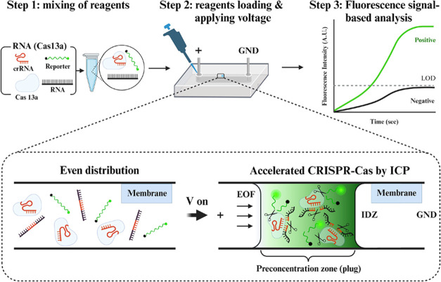
It is extremely advantageous to detect nucleic acid levels in the early phases of disease management; such early detection facilitates timely treatment, and it can prevent altogether certain cancers and infectious diseases. A simple, rapid, and versatile detection platform without enzymatic amplification for both short and long sequences would be highly desirable in this regard. Our study addresses this need by introducing IMACC, an ICP-based Microfluidic Accelerator Combined with CRISPR, for amplification-free nucleic acid detection. It exploits electrokinetically induced ion concentration polarization (ICP) to concentrate target nucleic acids and CRISPR reagents near the depletion zone boundary within a microfluidic channel. This localized accumulation accelerates the CRISPR-guided promiscuous cleavage of reporter molecules while enhancing their fluorescence signals simultaneously. Simultaneous accumulation of RNA and ribonucleoproteins (RNP) in confined spaces was validated experimentally and numerically, showing overlapping regions. IMACC enabled detection of miRNA-21 (22 bp) down to 10 pM within 2 min of ICP. IMACC ensured CRISPR specificity (single mismatch (N = 1) sensitivity) during ICP, as shown by off-target and mismatch sequence experiments. IMACC was applied to long RNA samples (i.e., SARS-CoV-2), but it statistically remained challenging at this point due to nonlinear intensity trends with copy numbers and large deviations. IMACC enabled rapid detection of short RNAs such as microRNAs using only basic CRISPR reagents in a single microfluidic channel, eliminating the need for extra enzymes or buffer sets, streamlining workflow and reducing turnaround time. IMACC has the potential to advance CRISPR diagnostics and holds promise for improved detection and future prescreening applications.


Conflict of interest statement

The authors declare no competing financial interest.

Figures

Figure 1
Figure 1
Detection of RNA using an ICP-based Microfluidic Accelerator Combined with CRISPR (IMACC). The IMACC workflow consists of three key steps: (1) Reagent Preparation: Cas13a, crRNA, reaction buffers, and target RNA are mixed. (2) Loading and ICP generation: the reagent mixture is loaded into the reservoir, and a voltage is applied across the microfluidic channel. This generates a preconcentration zone, visible as a focused plug near the IDZ, where CRISPR collateral cleavage is accelerated due to a shorter diffusion length, while cleaved fluorescence reporters accumulate inside the plug. (3) Detection and Analysis: the fluorescence signals of cleaved reporters are monitored and analyzed.
Figure 2
Figure 2
Microfluidic accelerator with PolyAMPS hydrogel-based ion-selective membrane and detection of RNA (e.g., miRNA-21) in IMACC. (A) A photograph showing a single and straight microfluidic channel of the microfluidic accelerator and an enlarged bright-field image showing a PolyAMPS hydrogel-based ion-selective membrane polymerized in the middle of the channel. (B) Fluorescence signal generated from FAM dye as a function of preconcentration time t, which was cleaved by Cas13a-crRNA complex (RNP) targeting miRNA-21 at c = 100 nM in IMACC at 60 V. At t = 10 s, the fluorescence signal became visible as a plug that slowly receded to the anodic reservoir at t = 20 and 30 s while its signal intensity continuously increased. The scale bar is 200 μm. (C) Validation of the overlapping location of each CRISPR reagent during detection of miRNA-21 via IMACC. The fluorescence image showed the location of each CRISPR reagent tagged with three fluorescence dyes: Ribonucleoproteins (RNP), which is a complex form of Cas13a-crRNA was observed as red color due to Alexa Fluor 647 tagged on crRNA, and the cleaved reporter was observed as green due to the FAM tag on the reporter, and the miRNA-21 was observed as orange due to the TAMRA tagged on miRNA-21. There was approximately a t = 1 s difference in the observation time for each CRISPR reagent due to the plug’s receding during the ICP and the time lag by the fluorescence filter change. The scale bar (white) is 200 μm. (D) A set of images shown on the left shows numerically studied preconcentration plugs of Cas-crRNA complex (RNP) and RNAs during ICP, and their merged 2D image with mesh, shown on the right, demonstrates overlapping of each plug between Cas-crRNA (RNP: green) and RNAs (purple), which is comparable with the result from experiments. MEM stands for a cation-selective membrane. Scale bar (black): 20 units of the x-axis equals 400 μm.
Figure 3
Figure 3
Quantitative analysis for detection of the synthetic miRNA- 21 using various concentrations from c = 10 to 105 pM by IMACC at 60 V. The maximum intensity showed an increase as a function of the concentration of miRNA-21. All five concentrations showed maximum intensity values above the value of the noise level (control average +3σ: red dashed line).
Figure 4
Figure 4
Investigation specificity of IMACC using off-target or mismatch sequences. (A) The maximum intensity obtained from all the off-targets (miRNA-134, 155, and 483: black bar), and from (B) different numbers of mismatches (N = 1, 2, and 3: black bar) were much less than that of the target (miRNA-21: blue bar), demonstrating specific detection of IMACC.

Similar articles

References

    1. Calin G. A.; Croce C. M. MicroRNA signatures in human cancers. Nat. Rev. Cancer 2006, 6 (11), 857–866. 10.1038/nrc1997. - DOI - PubMed
    2. Grubaugh N. D.; Ladner J. T.; Lemey P.; Pybus O. G.; Rambaut A.; Holmes E. C.; Andersen K. G. Tracking virus outbreaks in the twenty-first century. Nat. Microbiol. 2019, 4 (1), 10–19. 10.1038/s41564-018-0296-2. - DOI - PMC - PubMed
    3. Siravegna G.; Marsoni S.; Siena S.; Bardelli A. Integrating liquid biopsies into the management of cancer. Nat. Rev. Clin. Oncol. 2017, 14 (9), 531–548. 10.1038/nrclinonc.2017.14. - DOI - PubMed
    1. Yang S.; Rothman R. E. PCR-based diagnostics for infectious diseases: uses, limitations, and future applications in acute-care settings. Lancet Infect. Dis. 2004, 4 (6), 337–348. 10.1016/S1473-3099(04)01044-8. - DOI - PMC - PubMed
    2. Burki T. K. Testing for COVID-19. Lancet Respir. Med. 2020, 8 (7), E63–E64. 10.1016/S2213-2600(20)30247-2. - DOI - PMC - PubMed
    3. Weissleder R.; Lee H.; Ko J.; Pittet M. J. COVID-19 diagnostics in context. Sci. Transl. Med. 2020, 12 (546), eabc193110.1126/scitranslmed.abc1931. - DOI - PubMed
    1. Mahony J. B.; Blackhouse G.; Babwah J.; Smieja M.; Buracond S.; Chong S.; Ciccotelli W.; O’Shea T.; O’Shea T.; Alnakhli D.; Griffiths-Turner M. Cost Analysis of Multiplex PCR Testing for Diagnosing Respiratory Virus Infections. J. Clin. Microbiol. 2009, 47 (9), 2812–2817. 10.1128/JCM.00556-09. - DOI - PMC - PubMed
    1. Wang D. G.; Brewster J. D.; Paul M.; Tomasula P. M. Two Methods for Increased Specificity and Sensitivity in Loop-Mediated Isothermal Amplification. Molecules 2015, 20 (4), 6048–6059. 10.3390/molecules20046048. - DOI - PMC - PubMed
    2. Phillips E. A.; Moehling T. J.; Bhadra S.; Ellington A. D.; Linnes J. C. Strand Displacement Probes Combined with Isothermal Nucleic Acid Amplification for Instrument-Free Detection from Complex Samples. Anal. Chem. 2018, 90 (11), 6580–6586. 10.1021/acs.analchem.8b00269. - DOI - PMC - PubMed
    1. Yozwiak N. L.; Skewes-Cox P.; Stenglein M. D.; Balmaseda A.; Harris E.; DeRisi J. L. Virus Identification in Unknown Tropical Febrile Illness Cases Using Deep Sequencing. PLoS Neglect. Trop. Dis. 2012, 6 (2), e148510.1371/journal.pntd.0001485. - DOI - PMC - PubMed
    2. Wilson M. R.; Naccache S. N.; Samayoa E.; Biagtan M.; Bashir H.; Yu G. X.; Salamat S. M.; Somasekar S.; Federman S.; Miller S.; et al. Actionable Diagnosis of Neuroleptospirosis by Next-Generation Sequencing. New Engl. J. Med. 2014, 370 (25), 2408–2417. 10.1056/NEJMoa1401268. - DOI - PMC - PubMed
    3. Langelier C.; Zinter M. S.; Kalantar K.; Yanik G. A.; Christenson S.; O’Donovan B.; White C.; Wilson M.; Sapru A.; Dvorak C. C.; et al. Metagenomic Sequencing Detects Respiratory Pathogens in Hematopoietic Cellular Transplant Patients. Am. J. Respir. Crit. Care 2018, 197 (4), 524–528. 10.1164/rccm.201706-1097LE. - DOI - PMC - PubMed

LinkOut - more resources