. 2025 Jun 3;13(6):e0284824.
doi: 10.1128/spectrum.02848-24. Epub 2025 Apr 30.

Inhibitory effect of plant flavonoid cyanidin on oral microbial biofilm

Affiliations

Inhibitory effect of plant flavonoid cyanidin on oral microbial biofilm

Lucille Rudin et al. Microbiol Spectr. .

Abstract

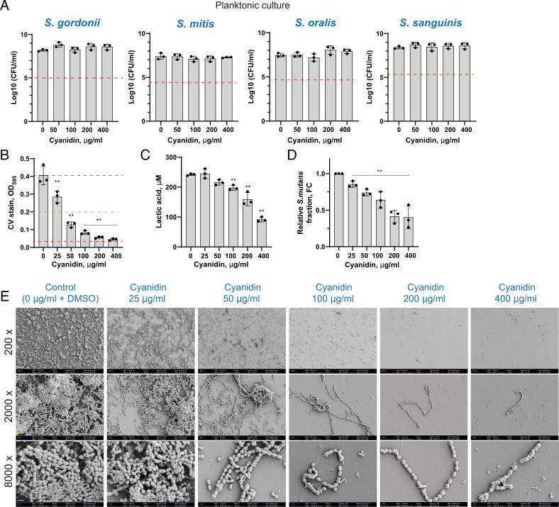
As primary colonizers of the tooth surface, oral streptococci play a crucial role in dental caries development. Numerous natural compounds, including flavonoids, are emerging as promising agents for inhibiting dental biofilm formation without compromising bacterial viability, underscoring their potential in non-bactericidal antibiofilm strategies. This study investigated the effects and mechanism of action of the unmodified plant flavonoid cyanidin on the growth and sucrose-dependent biofilm formation of oral streptococci, with a particular focus on the cariogenic pathogen Streptococcus mutans. At concentrations above 100 µg/mL, cyanidin significantly inhibited biofilm formation by S. mutans without impacting bacterial viability. The flavonoid reduced the biomass of surface-associated bacteria and exopolysaccharides (EPS), particularly by inhibiting water-insoluble glucan (WIG) production mediated by the glucosyltransferases GtfB and GtfC. While cyanidin did not exhibit a bactericidal effect on early colonizer streptococci, such as Streptococcus sanguinis, Streptococcus gordonii, Streptococcus oralis, and Streptococcus mitis, it showed a significant inhibitory effect on bacterial acidogenicity and mixed-species streptococcal biofilms in the presence of S. mutans. Remarkably, cyanidin gradually reduced the proportion of S. mutans in the mixed biofilm, suggesting a selective impact that may promote a more commensal-dominant community by disrupting S. mutans glucan production and biofilm competitiveness.

Importance: The identification of compounds with potent antibiofilm effects that do not compromise bacterial viability presents a promising strategy for oral health management. By preventing biofilm formation and keeping bacteria in a planktonic state, such agents could enhance bacterial susceptibility to targeted therapies, including probiotics or phage-based treatments. Cyanidin, which exhibits strong antibiofilm activity against oral streptococcal biofilms, reduces bacterial acidogenicity and may promote a more commensal-dominant biofilm in vitro, potentially hindering the maturation of cariogenic biofilms.

Keywords: Streptococcus mutans; biofilms; dental plaque; flavonoids.


Conflict of interest statement

The authors declare no conflict of interest.

Figures

Fig 1
Fig 1
Antibiofilm activity of cyanidin against S. mutans. (A) Chemical structure of cyanidin with PubChem ID indicated. (B) The effect of cyanidin on 24 h biofilms of S. mutans formed on a 96-well plate in brain heart infusion broth supplemented with 1% sucrose (BHIS). Biofilms were quantified by crystal violet staining and measuring absorbance at 595 nm. Green, orange, and red dashed lines indicate 0%, 50%, and 90% of biofilm inhibition, respectively. (C) Production of lactic acid by S. mutans treated with 50, 100, 200, and 400 µg/mL of cyanidin. (D and E) The effect of cyanidin on S. mutans cell viability growing for 24 h in planktonic culture (D) or collected all together after the biofilm assay (E). After the incubation, cell suspensions were serially diluted and plated for CFU enumeration. Red dashed lines indicate a 3-log10 reduction (99.9% reduction in CFU/mL) considered bactericidal. (F) Representative image from scanning electron microscopy (SEM) analysis of S. mutans biofilm formed on borosilicate glass disks submerged in a 96-well plate for biofilm assay. Disks were observed at magnifications of 200×, 2,000×, and 8,000× in three arbitrarily selected locations. The yellow scale bar represents 40, 4, and 1 µm for respective magnifications. (G) The proportion of live/dead cells was calculated from the cell biomasses reconstructed from CLSM images. “Live” cell fraction corresponds to the SYTO9-stained cells, while “dead” corresponds to the PI-stained cells. Chlorhexidine, 0.02%, was used as a bactericidal control. (H) Representative images of the double-labeled 24 h biofilms treated with cyanidin. Bacterial cells are shown in green, and exopolysaccharide (EPS) is in red. Three-dimensional reconstructions based on fluorescent intensity values were performed with Imaris 9.0.0. (I) Bacterial and EPS biomasses as calculated by Imaris per observational area after the three-dimensional reconstructions. (J) The effect of cyanidin on 24 h biofilms of S. mutans formed in the presence of 0.1%, 0.2%, and 0.5% sucrose. For each condition, CV staining values have been normalized to the non-treated control. Green, orange, and red dashed lines indicate 0%, 50%, and 90% of biofilm inhibition, respectively. (K) The effect of cyanidin on water-soluble and water-insoluble glucans produced by S. mutans cells in a 24 h biofilm assay, as measured by the anthrone-sulfuric method. (L) The effect of 200 µg/mL cyanidin on the biofilm-associated gene expression levels in a 24 h biofilm. The relative expression levels were quantified by real-time PCR with 16S rRNA as an internal control. (M) The best docked interaction of cyanidin with the active sites of GtfB (green) and GtfC (gray) of S. mutans. The cyanidin molecule is shown in cyan, and hydrogen bonds with displayed amino acids are shown as dashed yellow lines. (N) The effect of cyanidin-3-O-glucoside, proanthocyanidin B2, and quercetin on 24 h biofilms of S. mutans formed on a 96-well plate in brain heart infusion broth supplemented with 1% sucrose (BHIS). In all panels, bacterial culture supplemented with 5% dimethyl sulfoxide (DMSO) was used as a control. Bars represent the mean of at least three biological replicates. Error bars show standard deviation. **P < 0.0001.
Fig 2
Fig 2
Cyanidin effect on commensal oral streptococci. (A) The effect of cyanidin on oral streptococci cell viability growing for 24 h in planktonic culture. After the incubation, cell suspensions were serially diluted and plated for CFU enumeration. Red dashed lines indicate a 3-log10 reduction (99.9% reduction in CFU/mL) considered bactericidal. (B) The effect of cyanidin on a 24 h mixed streptococcal biofilm (S. mutans together with commensal streptococci such as S. sanguinis, S. mitis, S. oralis, and S. gordonii) formed on a 96-well plate in brain heart infusion broth supplemented with 1% sucrose (BHIS). Biofilms were quantified by crystal violet staining and measuring absorbance at 595 nm. Green, orange, and red dashed lines indicate 0%, 50%, and 90% of biofilm inhibition, respectively. (C) Production of lactic acid by streptococcal mixture treated with 25, 50, 100, 200, and 400 µg/mL of cyanidin. (D) The fold change values of the S. mutans fraction in treated mixed biofilms relative to untreated controls, calculated using the ΔΔCt method. In all panels, bacterial culture supplemented with 5% DMSO was used as a control. Bars represent the mean of at least three biological replicates. Error bars show standard deviation. **P < 0.0001. (E) Representative image from SEM analysis of mixed streptococcal biofilms formed on borosilicate glass disks submerged in a 96-well plate for biofilm assay. Disks were observed at magnifications of 200×, 2,000×, and 8,000× in three arbitrarily selected locations. The yellow scale bar represents 40, 4, and 1 µm for respective magnifications.

Similar articles

References

    1. Flemming H-C, Wingender J, Szewzyk U, Steinberg P, Rice SA, Kjelleberg S. 2016. Biofilms: an emergent form of bacterial life. Nat Rev Microbiol 14:563–575. doi:10.1038/nrmicro.2016.94 - DOI - PubMed
    1. Mira A, Simon-Soro A, Curtis MA. 2017. Role of microbial communities in the pathogenesis of periodontal diseases and caries. J Clin Periodontol 44:S23–S38. doi:10.1111/jcpe.12671 - DOI - PubMed
    1. Lemos JA, Palmer SR, Zeng L, Wen ZT, Kajfasz JK, Freires IA, Abranches J, Brady LJ. 2019. The biology of Streptococcus mutans. Microbiol Spectr 7. doi:10.1128/microbiolspec.GPP3-0051-2018 - DOI - PMC - PubMed
    1. Willenborg J, Goethe R. 2016. Metabolic traits of pathogenic streptococci. FEBS Lett 590:3905–3919. doi:10.1002/1873-3468.12317 - DOI - PubMed
    1. Paes Leme AF, Koo H, Bellato CM, Bedi G, Cury JA. 2006. The role of sucrose in cariogenic dental biofilm formation—new insight. J Dent Res 85:878–887. doi:10.1177/154405910608501002 - DOI - PMC - PubMed

MeSH terms

LinkOut - more resources