. 2025 May 20;99(5):e0024025.
doi: 10.1128/jvi.00240-25. Epub 2025 Apr 30.

Novel murine model of human astrovirus infection reveals cardiovascular tropism

Affiliations

Novel murine model of human astrovirus infection reveals cardiovascular tropism

Macee C Owen et al. J Virol. .

Abstract

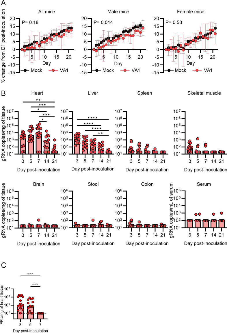
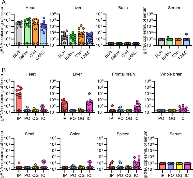
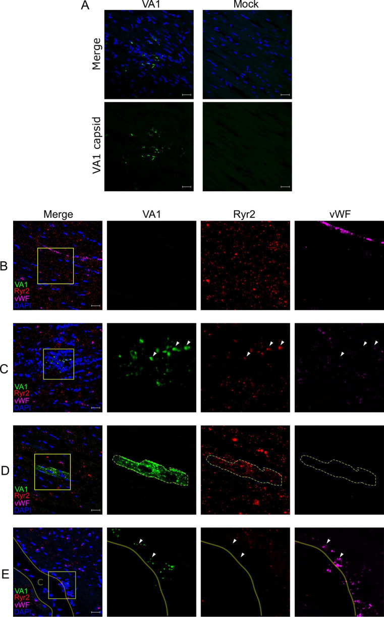
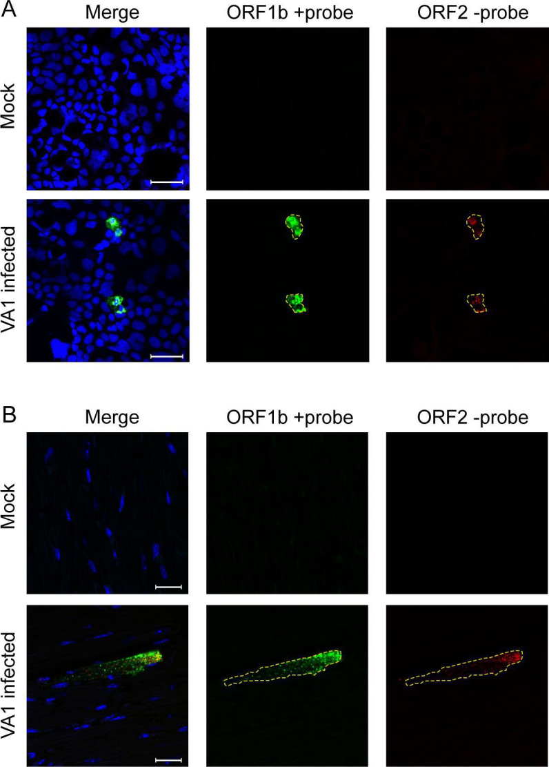
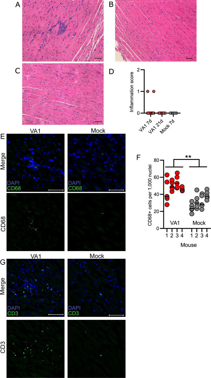
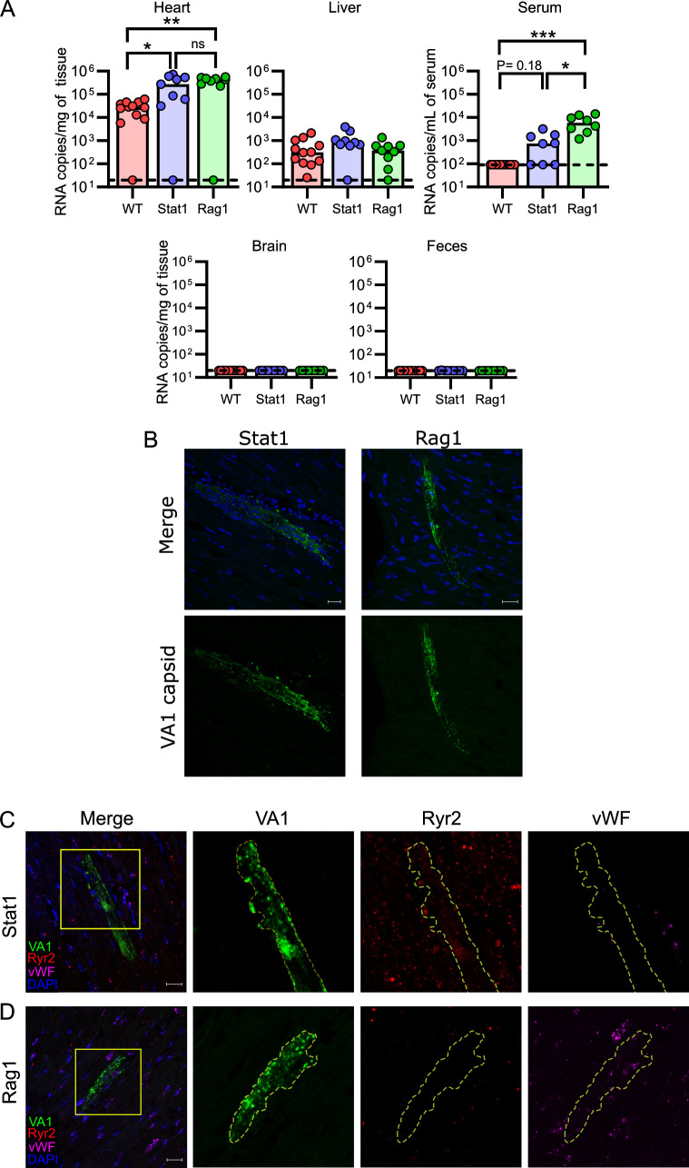
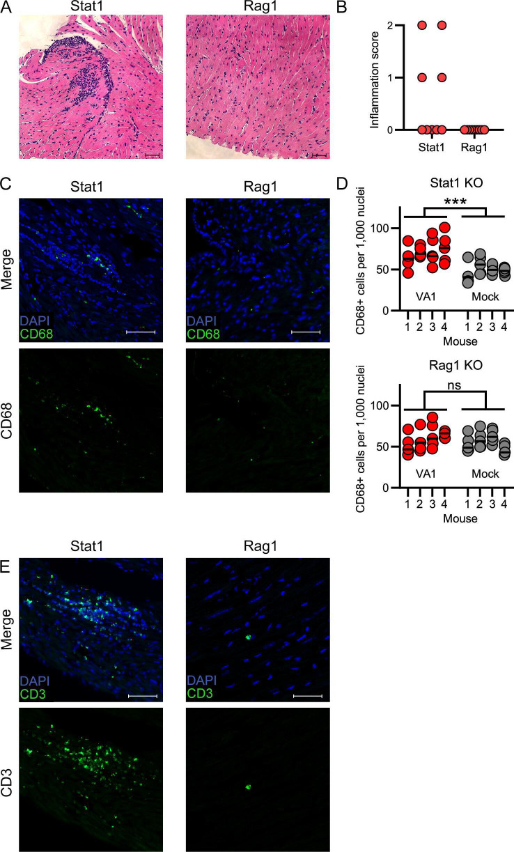
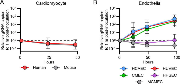
Astroviruses are a common cause of gastrointestinal disease in humans and have been linked to fatal cases of encephalitis. A major barrier to the study of human-infecting astroviruses is the lack of an in vivo model as previous attempts failed to identify a host that supports viral replication. We describe a novel murine model of infection using astrovirus VA1/HMO-C (VA1), an astrovirus with high seroprevalence in humans. VA1 is cardiotropic, and viral RNA levels peak in the heart tissue 7 days post-inoculation in multiple different murine genetic backgrounds. Infectious VA1 particles could be recovered from heart tissue 3 and 5 days post-inoculation. Viral capsid was detected intracellularly in the heart tissue by immunostaining, and viral RNA was detected in cardiac myocytes, endocardium, and endothelial cells based on fluorescent in situ hybridization and confocal microscopy. Histologically, we identified inflammatory infiltrates consistent with myocarditis in some mice, with viral RNA colocalizing with the infiltrates. These foci contained CD3 +T cells and CD68 +macrophages. Viral RNA levels increased by >10 fold in the heart tissue or serum samples from Rag1 or Stat1 knockout mice, demonstrating the role of both adaptive and innate immunity in the response to VA1 infection. Based on the in vivo tropisms, we tested cardiac-derived primary cells and determined that VA1 can replicate in primary human cardiac endothelial cells, suggesting a novel cardiovascular tropism in human cells. This novel in vivo model of a human-infecting astrovirus enables further characterization of the host immune response and reveals a new cardiovascular tropism of astroviruses.

Importance: Astroviruses routinely cause infections in humans; however, few methods were available to study these viruses. Here, we describe the first animal system to study human-infecting astroviruses by using mice. We demonstrate that mice are susceptible to astrovirus VA1, a strain that commonly infects humans and has been linked to fatal brain infections. The virus infects the heart tissue and is associated with inflammation. When mice with impaired immune systems were infected with VA1, they were found to have higher amounts of the virus in their hearts and blood. We found that VA1 can infect cells from human blood vessels of the heart, which is associated with human health. This model will enable us to better understand how astroviruses cause disease and how the immune system responds to infection. Our findings also suggest that astroviruses could be linked to cardiovascular diseases, including in humans.

Keywords: animal models; astrovirus; myocarditis; tropism; viral immunity; virology.


Conflict of interest statement

The authors declare no conflict of interest.

Figures

Fig 1
Fig 1
Inoculation of VA1 in wild-type A/J mice results in accumulation of viral RNA in the heart tissue. Wild-type A/J mice were intraperitoneally inoculated with VA1. (A) Male but not female mice had reduced weight gain following inoculation with VA1 compared to sex-matched mock-infected mice. (B) Over the course of 21 days post-inoculation, the viral RNA load was measured from tissues. The highest viral RNA loads were in the heart, peaking 7 days post-inoculation but remained detectable in some mice at day 21. Viral RNA was also detected from the liver, spleen, and skeletal muscle, while other tissues had sporadic detection of VA1 RNA. The dashed line indicates the limit of detection. (C) Viral infectious titers from heart homogenates collected from VA1-inoculated mice were measured by a focus-forming assay. Infectious virus could be detected 3 and 5 days post-inoculation, while it was undetectable on day 7. The dashed line represents the limit of detection. For (B) and (C), * denotes P < 0.05, **P ≤ 0.01, and ***P ≤ 0.001.
Fig 2
Fig 2
VA1 is cardiotropic in other mouse strains, while inoculation by other routes does not result in significant cardiac infection. Commonly studied mouse strains (C57BL/6 [BL/6], Balb/c, C3H/HeJ, and J:ARC Swiss Outbred) were intraperitoneally inoculated with VA1, and viral load was measured by qRT-PCR 7 days post-inoculation (A) Like A/J mice, other strains of mice have a significant viral load in the heart and liver tissue, while there was only sporadic detection in the serum and brain tissue. (B) Different routes of inoculation were tested in wild-type A/J mice, including intraperitoneal (IP; data from Fig. 1B, per os (PO), oral gavage (OG), and intracranial (IC) inoculation. Viral RNA loads were measured 7 days after inoculation. PO and OG inoculation did not result in significant infection of mice, including in the heart tissue compared to IP inoculation. IC inoculation resulted in detection of low quantities of viral RNA from the brain tissue, including whole or frontal brain tissue, but was several logs lower than what can be detected in the heart tissue from IP inoculation. Viral RNA was also detected from the liver and spleen, while low quantities were detected from the heart. For all graphs, the dashed line is the limit of detection.
Fig 3
Fig 3
VA1 RNA intracellularly localizes to cardiac myocytes and endocardial cells in wild-type A/J mice. (A) Immunofluorescence staining of VA1- or mock-inoculated mouse hearts. Tissues were processed for immunofluorescence and stained with an antibody to the VA1 capsid (green) and counterstained with DAPI. VA1-infected mice had positive punctate signals in regions with increased nuclei. No staining was observed in mock-inoculated mice. (B–E) A fluorescent in situ hybridization (FISH) assay using probes specific to VA1 open reading frame 2 (ORF2; green) was developed and imaged by confocal microscopy. After 7 days post-inoculation, the heart tissue was obtained and stained with probes to VA1 and to host markers for cardiac cell types including cardiomyocytes (Ryr2 probe; red) and endothelial cells (vWF probe; magenta). Merged fluorescence images are shown with counterstaining of nuclei (performed with DAPI). Boxes demonstrate areas of interest that were further magnified to demonstrate the signal from individual fluorescent channels. Outlines indicate cellular borders of cells containing VA1 RNA. White triangles highlight further areas of staining by the VA1 and host marker probes (B) No staining for VA1 was detected in mock-infected mouse hearts. (C–E) VA1-infected hearts stain positive for detectable VA1 colocalizing with (C) within foci of dense cellular infiltrates and colocalizing with Ryr2, (D) cardiomyocytes expressing Ryr2 without cellular infiltrates, and (E) within a region of endocardial cells expressing vWF. The solid line and the label C denote the open space of the heart chamber. Scale bars represent 20 µm.
Fig 4
Fig 4
Detection of negative-strand RNA from heart tissue (A) Strand-specific FISH staining of Caco-2 cells that were either mock-inoculated or -infected with VA1. The cells were stained with positive and negative sense probes to VA1 48 hours after inoculation. Both RNA strands were detected only in VA1-inoculated cells, consistent with infection and active replication. (B) Mock- or VA1-inoculated mice were stained with the strand-specific FISH probes. VA1-inoculated mouse hearts had both positive and negative sense strands being detected, demonstrating that active viral replication was occurring in cardiac tissue. Scale bars represent 20 µm.
Fig 5
Fig 5
Histological findings consistent with myocarditis are present in VA1-infected heart tissue. Heart tissue histology was analyzed by hematoxylin and eosin staining, with representative images shown. (A) Inflammatory infiltrates were present in some heart tissue on day 7, but (B) absent at day 21 or (C) in mock-inoculated mouse hearts. Scale bars represent 50 µm. (D) The presence of focal infiltrates was scored, and they were only present in hearts on day 7 post-inoculation. (E) Immunofluorescence was used to stain tissues for CD68, a marker of macrophages and other monocytes. Cells comprising the cellular infiltrates present in VA1-infected heart tissue were CD68-positive. Scale bars represent 20 µm. (F) CD68-positive cells were counted in representative tissue regions that lacked foci of cellular infiltrates. A significant increase in CD68-positive cells was identified in VA1-infected (mean 48.5 CD68 +cells per 1,000 nuclei) compared to mock-infected hearts (mean 31.64 CD68 +cells per 1,000 nuclei; nested T test t (6)= 4.35 P = 0.005). ** represents P ≤ 0.01. A total of four mice in each group were analyzed with five representative sections from each mouse used for counting per mouse. Horizontal lines represent the median value. (G) Immunofluorescent staining for CD3, a marker of T cells. As with CD68, many of the cells with a focus of infiltrating cells were CD3-positive and were absent in mock-infected mice. Scale bars are 20 µm.
Fig 6
Fig 6
Immunodeficient mice support higher loads of VA1 in the heart and serum. Wild-type (WT) C57BL/6, Rag1 KO, and Stat1 KO mice were inoculated with VA1 by the IP route, and tissues were collected 7 days post-inoculation. (A) VA1 copy number detected by qRT-PCR revealed higher quantities of VA1 RNA from the heart tissue and serum of Rag1 KO mice compared to C57BL/6 WT (WT data from Fig. 2). Stat1 KO mice had higher quantities of viral RNA in the heart tissue. *** represents P value ≤ 0.001, ** represents P value ≤ 0.01, * represents P value ≤ 0.05, and ns represents P value > 0.05. (B) Immunofluorescence for the VA1 capsid in immunodeficient mouse hearts. Both genetic backgrounds had positive staining for the viral capsid, including large cell(s) with morphology consistent with that of cardiac myocytes. (C–D) Representative fluorescent in situ hybridizations for VA1 (green) in the heart tissue, co-stained for cardiomyocytes (Ryr2 probe; red), and endothelial cells (vWF probe; magenta), with nuclei stained with DAPI (blue). Boxes highlight regions of interest that were further magnified. Scale bars represent 20 µm. Outlines highlight cells in which host markers localize to VA1 probes. (C) We detected VA1 RNA in cells expressing Ryr2, demonstrating infection of cardiac myocytes in both Stat1 and Rag1 mice, with a representative infection depicted from a Stat1 mouse. (D) In Rag1 KO mice, we also identified VA1-infected cells (green) that were expressing vWF (magenta) but were not expressing Ryr2 (red), suggestive of endothelial cells.
Fig 7
Fig 7
CD68- and CD3-positive cellular infiltrates are present in Stat1 KO but not Rag1 KO mice (A–B) Hematoxylin and eosin staining of heart tissue collected from (A) Stat1 KO mice shows clusters of cellular infiltration, which are absent in Rag1 KO mice. Scale bars represent 50 µm. (B) Quantification of inflammation scores from Stat1 KO and Rag1 KO heart tissue 7 days post-inoculation. Foci were identified in Stat1 KO mice, but none were present in Rag1 KO mice. (C) CD68-positive cellular infiltrates are present in Stat1 KO but not Rag1 KO mice, as detected by immunofluorescence. Scale bars represent 50 µm (D) In representative tissue regions excluding foci of infiltrating cells, Stat1 (nested T test t (6)= 5.65 P< 0.001) but not Rag1 KO mice (nested T test t (6)= 0.85 P = 0.43) had significant increases in the number of CD68-positive cells compared to mock-infected mice. Four mice per group were analyzed with five sections counted per mouse. Horizontal lines represent median value. *** represents P value ≤ 0.001, ns represents P value > 0.05. (E) Immunofluorescence of heart tissue for CD3 with a significant number of cells positive for CD3 in the infiltrating foci in Stat1 KO mice. Rare CD3 signal was detected in Rag1 KO mice. Scale bars represent 50 µm.
Fig 8
Fig 8
VA1 infects human endothelial-derived cell lines in vitro. Growth curves of VA1 using a multiplicity of infection of 3 in (A) primary human and mouse cardiac myocytes and (B) primary human endothelial cells including microvascular endothelial cells (CMECs), human coronary artery endothelial cells (HCAECs), human umbilical vein endothelial cells (HUVECs), human hepatic sinusoidal endothelial cells (HHSECs), and mouse cardiac endothelial cells (MCMECs). Each data point is normalized to the gRNA copy number present at 1 hour post-inoculation for each cell line, and geometric means are plotted, with error bars representing one geometric standard deviation. The horizontal dashed line represents the relative gRNA copy number at 1 hour post-inoculation.

Similar articles

References

    1. Janowski AB. 2021. Beyond the gastrointestinal tract: the emerging and diverse tissue tropisms of astroviruses. Viruses 13:732. doi:10.3390/v13050732 - DOI - PMC - PubMed
    1. Appleton H, Higgins PG. 1975. Letter: viruses and gastroenteritis in infants. Lancet 1:1297. doi:10.1016/s0140-6736(75)92581-7 - DOI - PubMed
    1. Bosch A, Pintó RM, Guix S. 2014. Human astroviruses. Clin Microbiol Rev 27:1048–1074. doi:10.1128/CMR.00013-14 - DOI - PMC - PubMed
    1. Harbour DA, Ashley CR, Williams PD, Gruffydd-Jones TJ. 1987. Natural and experimental astrovirus infection of cats. Vet Rec 120:555–557. doi:10.1136/vr.120.23.555 - DOI - PubMed
    1. Kurtz JB, Lee TW, Craig JW, Reed SE. 1979. Astrovirus infection in volunteers. J Med Virol 3:221–230. doi:10.1002/jmv.1890030308 - DOI - PubMed

LinkOut - more resources