. 2025 May 22;207(5):e0050324.
doi: 10.1128/jb.00503-24. Epub 2025 Apr 30.

FleQ finetunes the expression of a subset of BrlR-activated genes to enable antibiotic tolerance by Pseudomonas aeruginosa biofilms

Affiliations

FleQ finetunes the expression of a subset of BrlR-activated genes to enable antibiotic tolerance by Pseudomonas aeruginosa biofilms

Victoria I Oladosu et al. J Bacteriol. .

Abstract

The transcriptional regulator FleQ contributes to Pseudomonas aeruginosa biofilm formation by activating the expression and biosynthesis of matrix exopolysaccharides in a manner dependent on c-di-GMP. However, little is known about the role of FleQ in the antibiotic tolerance phenotype of P. aeruginosa biofilms. Inactivation of fleQ impaired biofilm formation and rendered biofilms susceptible to tobramycin and norfloxacin. The phenotypes were similar to biofilms inactivated in sagS encoding the orphan sensor SagS that promotes the switch from planktonic to biofilm growth via BfiSR and antibiotic tolerance via BrlR. While FleQ was found to contribute to biofilm formation independently of SagS and BfiSR, FleQ instead converged with SagS-dependent regulation at the level of BrlR. This was supported by multicopy expression of sagS failing to restore biofilm antibiotic tolerance by ΔfleQ to wild-type levels (and vice versa) and by biofilms formed by the ΔfleQΔsagS double mutant being as susceptible as ΔfleQ and ΔsagS biofilms. Increased antibiotic susceptibility was independent of BrlR abundance or BrlR DNA binding but coincided with significantly reduced transcript abundance of the BrlR-activated mexCD-oprJ and PA1874-77, encoding an ABC transporter previously shown to contribute to the tolerance of biofilms to tobramycin and norfloxacin. FleQ- dependent regulation of gene expression was indirect. Co-immunoprecipitation and BACTH assays indicated FleQ to interact with SagS via its HisKA-Rec domain, likely suggesting FleQ and SagS to likely work in concert to enable biofilm antibiotic tolerance, by finetuning the expression of BrlR activated genes.IMPORTANCEIn P. aeruginosa, FleQ inversely regulates the expression of genes encoding flagella and biofilm matrix components, including exopolysaccharide (Pel, Psl) in a manner dependent on the levels of c-di-GMP. Our findings expand on the role of FleQ from regulating the transition to the biofilm mode of growth to FleQ contributing to the antimicrobial tolerance phenotype of biofilms, by FleQ affecting the expression of PA1874-77, a downstream target of the SagS-dependent transcriptional regulator BrlR. Importantly, our findings suggest FleQ works in concert with SagS, likely via FleQ-SagS protein-protein interactions, to enable the formation of inherently tolerant P. aeruginosa biofilms.

Keywords: BACTH; BrlR; DNA binding; biofilm-MBC; immunoblot; protein-protein interaction; pulldown; resistant to killing.


Conflict of interest statement

The authors declare no conflict of interest.

Figures

Fig 1
Fig 1
FleQ and SagS are both required for biofilm formation. Biofilms were grown in flow cells under flowing conditions for 6 days. Biofilms were stained using BacLight LIVE/DEAD prior to visualizing the biofilm architecture by confocal microscopy. (A) Representative confocal images of biofilms by P. aeruginosa PAO1, ΔfleQ and ΔfleQ/pMJT-fleQ. (B) Representative confocal images of ΔfleQ and ΔfleQ expressing sagS (ΔfleQ/pJN-sagS) and ΔsagS expressing fleQ (ΔsagS/pMJT-fleQ). White bar = 100 µm. All experiments were performed in triplicate. (C) Growth curves. P. aeruginosa PAO1, ΔfleQ, and ΔfleQ/pMJT-fleQ were grown in VBMM at 37°C and 220 rpm and the absorbance was determined at 600 nm. Experiments were carried out in triplicate. Error bars indicate standard deviations.
Fig 2
Fig 2
FleQ contributes to the susceptibility phenotype of P. aeruginosa biofilms. (A) Biofilms of indicated strains were grown in tube reactors under flowing conditions for 3 days and subsequently exposed to tobramycin (150 µg/mL) and norfloxacin (450 µg/mL) for 1 h under flowing conditions. Biofilm susceptibility was determined by log10 reduction. *, **, ***, ****, significantly different from the negative control (P < 0.05, P < 0.001, P < 0.0004, P < 0.0001, respectively) using ANOVA with Dunnett’s T3 multiple comparisons test. (B) P. aeruginosa PAO1 and ΔfleQ were grown planktonically to exponential phase and then exposed to tobramycin (50 µg/mL) and norfloxacin (50 µg/mL). Susceptibility was determined by log10reduction. ***, significantly different (P < 0.005) relative to PAO1, as determined using an unpaired t-test with Welch’s correction. ns, not significant. (C) Biofilm-MBC assays. P. aeruginosa PAO1, ΔfleQ, and ΔsagS were grown as biofilms for 3 days and subsequently treated for 24 h with tobramycin (300 µg/mL) under continuous flowing conditions before recovering and enumerating surviving cells. Biofilm susceptibility to tobramycin was determined by viable counts (biofilm CFU, obtained from biofilm tube reactors having an inner surface area of 25 cm2). #, no viable bacteria were detected. ****, significantly different from the untreated biofilm (P < 0.0001) using two-way ANOVA with Sidak’s multiple comparisons test. All experiments were carried out at least in triplicate. Error bars indicate standard deviations.
Fig 3
Fig 3
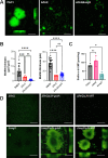
FleQ contributes to biofilm formation in a manner independent of SagS and SagS downstream signaling. (A) Representative confocal images of 3-day-old biofilms formed by P. aeruginosa PAO1, ΔfleQ, and double mutant ΔfleQΔsagS. White bar = 100 µm. (B) Quantitative of the biofilm biomass and biofilm height by COMSTAT of biofilms formed by P. aeruginosa PAO1, ΔfleQ, and double mutant ΔfleQΔsagS. **, ***, significantly different (P = 0.0001, <0.0001, respectively) relative to PAO1 using ANOVA followed by the Bartlett’s test. Ns, not significant. (C) Total cellular c-di-GMP levels in 6-day-old biofilms formed by P. aeruginosa PAO1, ΔfleQ, and ΔsagS, as determined by HPLC quantitative analysis, followed by normalization relative to total cell protein content. *, P < 0.05, relative to PAO1 using ANOVA followed by Dunnett’s T3 multiple comparisons test. (D) Representative confocal images of 6-day-old P. aeruginosa ΔfleQ and ΔsagS biofilms and mutant biofilms expressing bfiR and gcbA. gcbA encodes diguanylate cyclase GcbA, bfiR the two-component response regulator BfiR. White bar = 100 µm. All experiments were performed in triplicate. Error bars indicate standard deviations.
Fig 4
Fig 4
FleQ is required for the tolerance of biofilm cells to tobramycin in parallel with SagS. (A, B) Susceptibility phenotype of biofilms grown in tube reactors under flowing conditions for 3 days to tobramycin (150 µg/mL). Susceptibility was determined by log10 reduction. **, ***, ****, significantly different from PAO1 biofilms (P < 0.005, P < 0.001, P < 0.0001) using one-way ANOVA, followed by Tukey’s multiple comparison test. ns, not significant. (C) Representative immunoblot showing FleQ-dependent abundance of BrlR. Total cell extracts (TCE) obtained from biofilms of indicated P. aeruginosa strains expressing a chromosomally located V5/His6-tagged BrlR under the control of its own promoter were probed for the presence of BrlR by immunoblot analysis (IB) using anti-V5 antibodies (anti-V5). The corresponding SDS-PAGE gel image obtained post-transfer indicates equal loading. Representative images are shown. (D) Representative immunoblot showing FleQ-dependent DNA binding capability of BrlR. BrlR-DNA binding was determined using streptavidin magnetic bead binding assays. Binding assays were carried out using 5 pmol of BrlR-V5/His6 protein obtained from the indicated strains and 1 pmol biotinylated PbrlR. Non-biotinylated PbrlR (PbrlR-NB) was used as specific competitor DNA in 20-fold excess. BrlR binding to PbrlR was detected by immunoblot analysis using anti-V5 antibodies. +/−, indicates presence/absence of specific probe or competitor. All experiments were performed in triplicate. Error bars indicate standard deviations
Fig 5
Fig 5
FleQ indirectly affects the expression of a subset of BrlR target genes. (A) Transcript abundance of BrlR-target genes in biofilms formed by ΔfleQ and complemented ΔfleQ/pMJT-fleQ mutant strains relative to the wild type (PAO1). Quantitative RT-PCR (qRT-PCR) was carried out using RNA extracted from 3-day biofilms grown in fivefold diluted VBMM. arnC encodes a component required for resistance to cationic antimicrobial peptide such as colistin; mexA and mexC encode components of multi-drug efflux pumps; PA1874 belongs to an operon that encodes ABC transport systems; phoP and pmrA encode two-component response regulators that control the expression of the arn operon. cysD was used as the housekeeping gene. Experiments were carried out in triplicate. Error bars indicate standard deviations. *, ***, significantly different relative to the wild-type PAO1 (P < 0.5, P < 0.0001, respectively) using ordinary two-way ANOVA followed by Dunnett’s multiple comparison test. ns, not significant. (B, C) FleQ-DNA binding was determined using streptavidin magnetic bead binding assays, with FleQ binding to the respective promoter regions was detected by immunoblot analysis using anti-V5 antibodies, and subsequent analysis using ImageJ (48). Binding assays were carried out using 100 µg of total biofilm cell extract by PAO1 producing V5-tagged FleQ and 0–20 pmol biotinylated Ppel or PPA1874 or PmexC. (B) Quantitative analysis of FleQ DNA binding in the absence of additional c-di-GMP. (C) Representative immunoblots of FleQ DNA binding assays in the absence/presence of c-di-GMP and loading controls. Experiments were done in duplicate. Error bars indicate standard deviations.
Fig 6
Fig 6
FleQ interacts with SagS via its HisKA-Rec domain. (A) Representative image of immunoblot showing the abundance of FleQ in total cell extracts (TCE) and in pulldowns (IP) using SagS as prey. Strain ΔsagS/pMJT-1/pJN-sagS was used as control. M, protein ladder. (B) Representative image of immunoblot showing the abundance of SagS in total cell extracts (TCE) and in pulldowns (IP) using FleQ as prey. (C) Overview of SagS domains and SagS domain constructs. Lines underneath the domains indicate the composition of the SagS domain constructs, while the names of the resulting constructs are given next to the lines. (D) Representative images of bacterial adenylate cyclase two-hybrid assay (BACTH) results. DHM1/pUT18C-torS/pKT25-TorR was used as the positive control (+) and DHM1/pUT18C/pKT25 was used as the negative control (−). Bluish-green coloration represents a cleavage of X-gal by β-galactosidase indicative of protein-protein interaction. (E) Quantitative analysis of β-galactosidase activity using the Miller assay (51) of DHM1 strains co-expressing fleQ (pKT25-fleQ) and SagS-domain constructs (pUT18C -HisKA-Rec, pUT18C-HisKA, or pUT18C-HisKA-Rec). E. coli strains DHM1/pUT18C-torS/pKT25-torR and DHM1/pUT18C/pKT25 were used as controls. Experiments were done in triplicate. Error bars represent standard deviation. ** and ****, significantly different from the negative control (P < 0.01, P < 0.0001, respectively) using ANOVA followed by Dunnett’s T3 multiple comparisons test.
Fig 7
Fig 7
Model of FleQ finetuning BrlR target gene expression to modulate biofilm antibiotic tolerance. SagS indirectly contribute to the abundance and activation of the c-di-GMP responsive transcriptional regulator BrlR (36), likely by SagS contributing to the pool of available c-di-GMP generated by the diguanylate cyclase (DGC) PA3177 (33, 35). FleQ does not affect the abundance of BrlR or its DNA binding capability but indirectly affects the expression of a subset of BrlR target genes (17, 41). Dashed line, indirect effect of SagS on BrlR; dotted line, indirect effect of FleQ on BrlR, resulting in finetuning the expression of a subset of BrlR target genes.

Similar articles

References

    1. Sauer K, Stoodley P, Goeres DM, Hall-Stoodley L, Burmølle M, Stewart PS, Bjarnsholt T. 2022. The biofilm life cycle: expanding the conceptual model of biofilm formation. Nat Rev Microbiol 20:608–620. doi:10.1038/s41579-022-00767-0 - DOI - PMC - PubMed
    1. Davies D. 2003. Understanding biofilm resistance to antibacterial agents. Nat Rev Drug Discov 2:114–122. doi:10.1038/nrd1008 - DOI - PubMed
    1. Costerton JW, Cheng KJ, Geesey GG, Ladd TI, Nickel JC, Dasgupta M, Marrie TJ. 1987. Bacterial biofilms in nature and disease. Annu Rev Microbiol 41:435–464. doi:10.1146/annurev.mi.41.100187.002251 - DOI - PubMed
    1. Costerton JW, Stewart PS, Greenberg EP. 1999. Bacterial biofilms: a common cause of persistent infections. Science 284:1318–1322. doi:10.1126/science.284.5418.1318 - DOI - PubMed
    1. Ruiz M, Ewig S, Torres A, Arancibia F, Marco F, Mensa J, Sanchez M, Martinez JA. 1999. Severe community-acquired pneumonia. Risk factors and follow-up epidemiology. Am J Respir Crit Care Med 160:923–929. doi:10.1164/ajrccm.160.3.9901107 - DOI - PubMed

MeSH terms

LinkOut - more resources