. 2025 Jun 11;16(6):e0071925.
doi: 10.1128/mbio.00719-25. Epub 2025 Apr 30.

Stress-dependent activation of the Listeria monocytogenes virulence program ensures bacterial resilience during infection

Affiliations

Stress-dependent activation of the Listeria monocytogenes virulence program ensures bacterial resilience during infection

Mariya Lobanovska et al. mBio. .

Abstract

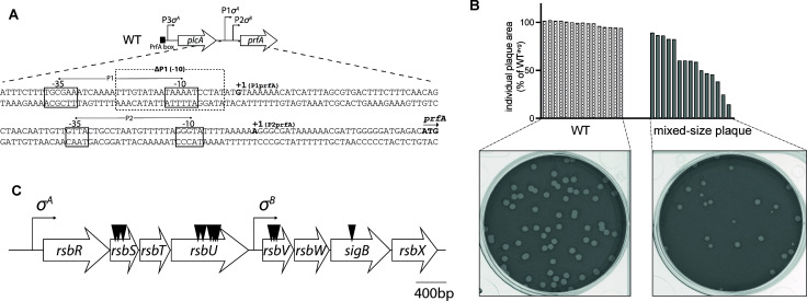
Listeria monocytogenes (Lm) is a Gram-positive, facultative intracellular pathogen that uses both a housekeeping (P1) and stress-activated (Sigma B-dependent) promoter (P2) to express the master virulence regulator PrfA. The Sigma B regulon contains over 300 genes known to respond to different stressors. However, the role of Sigma B in the regulation of prfA during the infection remains uncertain. To define pathways that lead to Sigma B-dependent prfA activation, we performed a genetic screen in L2 fibroblasts using ΔP1 Lm that only has the Sigma B-dependent promoter directly upstream of prfA. The screen identified transposon insertions in a large bacterial sensory organelle known as the stressosome. The absence of functional stressosome components resulted in heterogeneity within bacterial populations, with some bacteria behaving like wild type, while other members of the population exhibited defects in either vacuolar escape and/or cell-to-cell spread. We show that the heterogeneity of the stressosome mutants cannot be rescued by constitutive activation of PrfA. These data defined the importance of the stressosome in controlling bacterial homogeneity and characterized the function of the stressosome in robust virulence activation during infection. ΔP1 Lm model provides new opportunities to identify host-specific signals necessary for stressosome-dependent signaling during Listeria pathogenesis.IMPORTANCEMicrobial pathogens must adapt to varying levels of stress to survive. This study uncovered a link between stress sensing and activation of the virulence program in a facultative intracellular pathogen, Listeria monocytogenes. We show that host-imposed stress is sensed by the signaling machinery known as the stressosome to ensure robust and resilient virulence responses in vivo. Stressosome-dependent activation of the master virulence regulator PrfA was necessary to maintain L. monocytogenes homogeneity within the bacteria population during the transition between early and late stages of intracellular infection. This work also provides a model to further characterize how specific stress stimuli affect bacterial survival within the host, which is critical for our understanding of bacterial pathogenesis.

Keywords: intracellular bacteria; macrophages; pathogenesis; sigma factors; stress adaptation; virulence regulation.


Conflict of interest statement

D.A.P. has a financial interest in Laguna Biotherapeutics, and both he and the company could benefit from the commercialization of the results of this research.

Figures

Fig 1
Fig 1
Mixed-size plaque phenotype in a ΔP1 background. (A) Schematic of the ΔP1 Lm strain used in the genetic screen. The ΔP1 strain contains a deletion of the −10 sequence of the Sigma A-dependent P1 promoter. Physical map of prfA promoter region of Lm is adapted from Freitag and Portnoy (8). (B) Mixed-size plaque formation. Transposon mutants identified in the screen formed plaques of mixed sizes. The bar graph depicts plaque size morphology of a representative transposon mutant compared to WT plaques. (C) The stressosome locus. Eight-gene operon encoding different components of the stressosome signaling pathway contains a distal Sigma A-dependent promoter and Sigma B-dependent promoter positioned within the operon. Transposon insertion sites identified in the screen are shown by black arrowheads.
Fig 2
Fig 2
In vitro characterization of stressosome mutants. (A) Growth of the indicated strains at 37°C in BHI broth. Overnight bacterial cultures were normalized to an OD600 = 0.03 in fresh BHI and grown at 37°C for 15 h with agitation. The graph represents pooled data from three independent biological repeats. No statistical difference was detected between the strains. (B) Plaque area, presented as a percentage of WT, of indicated strains was measured following a 3-day infection of L2 cells. Data represent the mean of three independent experiments. (C and D) Growth curves in bone marrow-derived macrophages pre-treated with PAM3CK and infected with indicated strains at MOI of 0.25. Gentamicin (50 µg/mL) was added 1 h post-infection to kill extracellular bacteria. CFUs were counted at indicated time points, and the experiments were performed three times. (E) Quantification of cytosolic Lm expressed as a percentage of p62+ Lm+ bacteria. BMMs were infected with the indicated strains for 1.5 h. Cells were washed, fixed, and stained with anti-Listeria and anti-p62 antibodies. The percentage of Lm that escapes the phagosome and colocalizes with p62 is shown. At least 70 bacteria were analyzed per strain, and the experiment was performed two times. (F) Indicated strains were tested for sensitivity to the thiol-specific oxidant diamide by a disk diffusion assay. Zone of inhibition reflects the diameter presented as a percentage of WT. Data represent the mean of three independent experiments. For all experiments: Error bars indicate standard deviation. One-way ANOVA was used for multiple comparisons with WT (A–D, F) or WT and ΔP1 (E). ns, not significant, *P < 0.05; **P < 0.01; ***P < 0.001; ****P < 0.0001.
Fig 3
Fig 3
ΔP1 stressosome mutants have decreased PrfA expression. Indicated strains were grown to mid-log in iLSM + TCEP at 37°C, and samples were collected for RNA and Western blot analysis. (A) prfA expression was measured using qRT-PCR and normalized to 16S rRNA. One-way ANOVA was used to calculate for multiple comparisons with ΔP1. ns, not significant, *P < 0.05; **P < 0.01. (B) PrfA was detected using anti-PrfA antibody, and P60 expression was used as a control. The data represent three independent biological repeats. A ratio of PrfA/P60 was quantified using integrated density values.
Fig 4
Fig 4
Role of PrfA activation in the virulence of prfA promoter mutants in vivo. (A, B) Plaque formation of indicated strains measured in the L2 plaque assay. Plaque sizes are presented as a percentage of WT. Data are mean ± SD and represent three independent experiments. One-way ANOVA with multiple comparisons to WT was used to calculate P value. ns, not significant, *P < 0.05; ***P < 0.001. (C) CD-1 mice were infected with each strain. Spleens and livers were collected 48 h post-infection for CFU quantification. Solid lines indicate the median, and the data represent two pooled biological repeats with 7–9 mice per strain. L.o.d, limit of detection. One-way ANOVA, multiple comparisons to either WT or WT ΔgshF as indicated, was used to calculate the P values. ns, not significant, **P < 0.01; ***P < 0.001; ****P < 0.0001.
Fig 5
Fig 5
Analysis of the stressosome operon deletion mutants. (A) Plaque formation of WT and ΔP1 Δstressosome was measured in L2 cells and presented as percentage of WT. Data represent at least three independent experiments, and error bars indicate SD. P values were calculated using one-way ANOVA, multiple comparisons with WT. ns, not significant, ****P < 0.0001. (B) CD-1 mice were infected with WT and ΔP1 Δstressosome mutants, and CFUs were measured 48 h post-infection in spleens and livers. Solid lines indicate medians. Data represent two biological repeats combining 8–13 mice per strain. P values were calculated using one-way ANOVA, multiple comparisons with WT control strain. ns, not significant, *P < 0.05; ****P < 0.0001.
Fig 6
Fig 6
Characterization of RsbR paralog mutants in vivo. (A) Schematic representation of rsbR2–rsbR5 paralog loci in Lm. (B) Plaque assay of RsbR paralog and RsbR1 mutants in ΔP1 background. Plaque area is shown as a percentage of WT. Error bars indicate SD and one-way ANOVA, multiple comparisons with WT, was used to calculate P values. ns, not significant, **P < 0.01. (C) Virulence of RsbR paralog mutants in mice is presented as CFUs per mouse in spleens and livers after 48 h. Two biological repeats of 7–9 mice per strain. Solid lines represent the median. P values were calculated using one-way ANOVA, multiple comparisons with ΔP1. ns, not significant, *P < 0.05; **P < 0.01.

Similar articles

References

    1. Freitag NE, Port GC, Miner MD. 2009. Listeria monocytogenes - from saprophyte to intracellular pathogen. Nat Rev Microbiol 7:623–628. doi: 10.1038/nrmicro2171 - DOI - PMC - PubMed
    1. Radoshevich L, Cossart P. 2018. Listeria monocytogenes: towards a complete picture of its physiology and pathogenesis. Nat Rev Microbiol 16:32–46. doi: 10.1038/nrmicro.2017.126 - DOI - PubMed
    1. Pizarro-Cerdá J, Cossart P. 2018. Listeria monocytogenes: cell biology of invasion and intracellular growth. Microbiol Spectr 6:gpp3-0013-2018. doi: 10.1128/microbiolspec.gpp3-0013-2018 - DOI - PMC - PubMed
    1. Johansson J, Mandin P, Renzoni A, Chiaruttini C, Springer M, Cossart P. 2002. An RNA thermosensor controls expression of virulence genes in Listeria monocytogenes. Cell 110:551–561. doi: 10.1016/s0092-8674(02)00905-4 - DOI - PubMed
    1. Loh E, Dussurget O, Gripenland J, Vaitkevicius K, Tiensuu T, Mandin P, Repoila F, Buchrieser C, Cossart P, Johansson J. 2009. A trans-acting riboswitch controls expression of the virulence regulator PrfA in Listeria monocytogenes. Cell 139:770–779. doi: 10.1016/j.cell.2009.08.046 - DOI - PubMed

MeSH terms

LinkOut - more resources