. 2025 Oct;41(10):1729-1742.
doi: 10.1007/s12264-025-01397-1. Epub 2025 Apr 30.

The Glutamate-gated Chloride Channel Facilitates Sleep by Enhancing the Excitability of Two Pairs of Neurons in the Ventral Nerve Cord of Drosophila

Affiliations

The Glutamate-gated Chloride Channel Facilitates Sleep by Enhancing the Excitability of Two Pairs of Neurons in the Ventral Nerve Cord of Drosophila

Yaqian Fan et al. Neurosci Bull. 2025 Oct.

Abstract

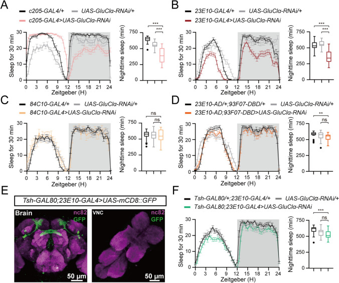
Sleep, an essential and evolutionarily conserved behavior, is regulated by numerous neurotransmitter systems. In mammals, glutamate serves as the wake-promoting signaling agent, whereas in Drosophila, it functions as the sleep-promoting signal. However, the precise molecular and cellular mechanisms through which glutamate promotes sleep remain elusive. Our study reveals that disruption of glutamate signaling significantly diminishes nocturnal sleep, and a neural cell-specific knockdown of the glutamate-gated chloride channel (GluClα) markedly reduces nocturnal sleep. We identified two pairs of neurons in the ventral nerve cord (VNC) that receive glutamate signaling input, and the GluClα derived from these neurons is crucial for sleep promotion. Furthermore, we demonstrated that GluClα mediates the glutamate-gated inhibitory input to these VNC neurons, thereby promoting sleep. Our findings elucidate that GluClα enhances nocturnal sleep by mediating the glutamate-gated inhibitory input to two pairs of VNC neurons, providing insights into the mechanism of sleep promotion in Drosophila.

Keywords: Drosophila; Glutamate-gated chloride channel; Neural activity; Neural circuit; Sleep; Ventral nerve cord.


Conflict of interest statement

Conflict of interest: The authors declare no competing financial interests.

Figures

Fig. 1
Fig. 1
GluClα expression in neurons is essential for nighttime sleep. A Sleep traces of control w;nSyb-GAL4/+ (black, n = 16), w;UAS-Dicer/+;UAS-vGluT-RNAi/+ (light gray, n = 27), w;UAS-Dicer/+;nSyb-GAL4/UAS-vGluT-RNAi (blue, n = 14), plotted as a 30-min moving average. B-E Total day sleep B, total nighttime sleep C, number of night sleep episodes D, and mean night sleep episode duration E for each genotype. F Total nighttime sleep of flies of each genotype with GluR knockdown using a nSyb-GAL4 driver and different UAS-GluR-RNAis. A single copy of the GAL4 driver was used for each RNAi line. G Sleep traces of control w;;nSyb-GAL4/+ (black, n = 50), w;UAS-GluClα-RNAi/+ (light gray, n = 52), w;UAS-GluClα-RNAi/+;nSyb-GAL4/+ (purple, n = 55) plotted as a 30-min moving average. H-K Total day sleep H, total nighttime sleep I, number of night sleep episodes J, and mean night sleep episode duration K for each genotype. For B, C, F, H, I, one-way ANOVA with Dunnett’s post hoc, ns, P > 0.05, ***P < 0.001; for D, E, J, K, Kruskal-Wallis followed by Dunn’s multiple comparison test, ns, P > 0.05, ***P < 0.001.
Fig. 2
Fig. 2
GluClα mutant flies exhibit reduced nighttime sleep. A Annotated transcription of the GluClα gene. Black triangle, the MI01156 insertion site; red square, the RNAi target site; black square, the point mutant site; dashed lines, the deleted regions of Df(3R)BSC636. The LexA insertion site is labeled above. B Sleep traces of control w1118 (black, n = 32), w;;GluClαglc1/+ (blue, n = 34), w;;GluClαMI01156/+(green, n = 30), w;;GluClαglc1/MI01156 (purple, n = 18), and w;;GluClαglc1/Df(3R)BSC636 (red, n = 21) plotted as 30-min moving averages. C-E Total nighttime sleep C, number of night sleep episodes D, and mean duration of night sleep episodes E for each genotype. In C, one-way ANOVA with Dunnett’s post hoc, ns, P > 0.05, ***P < 0.001; in D and E, Kruskal-Wallis followed by Dunn’s multiple comparisons test, ns, P > 0.05, ***P < 0.001.
Fig. 3
Fig. 3
Knockdown of GluClα in the dFB does not affect sleep. A Sleep traces and quantification of nighttime sleep of w;;c205-GAL4/+ (black, n = 30), w;UAS-GluClα-RNAi/+ (light gray, n = 29), and w;UAS-GluClα-RNAi/+;c205-GAL4/+ (light red, n = 49). B Sleep traces and quantification of nighttime sleep of w;;23E10-GAL4/+ (black, n = 31), w;UAS-GluClα-RNAi/+ (light gray, n = 29), and w;UAS-GluClα-RNAi/+;23E10-GAL4/+ (dark red, n = 56). C Sleep traces and quantification of nighttime sleep of w;;84C10-GAL4/+ (black, n = 42), w;UAS-GluClα-RNAi/+ (light gray, n = 51), and w;UAS-GluClα-RNAi/+;84C10-GAL4/+ (orange, n = 11). D Sleep traces and quantification of nighttime sleep of w;23E10-AD/+;97F07-DBD/+ (black, n = 31), w;UAS-GluClα-RNAi/+ (light gray, n = 29), and w;23E10-AD/UAS-GluClα-RNAi;97F07-DBD/+ (light orange, n = 23). E The brain and VNC of an adult Tsh-GAL80/+;23E10-GAL4/UAS-mCD8::GFP fly double-stained with anti-GFP (green) and anti nc82 (purple); scale bars, 50 µm. F Sleep traces and quantification of nighttime sleep of w;Tsh-GAL80/+;23E10-GAL4/+ (black, n = 50), w;UAS-GluClα-RNAi/+ (light gray, n = 52), and w;Tsh-GAL80/UAS-GluClα-RNAi;23E10-GAL4/+ (green, n = 55). For A, B, C, D, F, one-way ANOVA with Dunnett’s post hoc, ns, P > 0.05, ***P < 0.001.
Fig. 4
Fig. 4
Reduced sleep after GluClα knockdown in 23E10+ neurons maps to the VNC. A The brain and VNC of an adult w;vGluT-Trojan-GAL80/+;23E10-GAL4/UAS-mCD8::GFP fly double-stained with anti-GFP (green) and anti nc82 (purple); scale bars, 50 µm. B Sleep traces and quantification of nighttime sleep of w;vGluT-Trojan-GAL80;23E10-GAL4/+ (black, n = 45), w;UAS-GluClα-RNAi/+ (light gray, n = 31), and w;vGluT-Trojan-GAL80/UAS-GluClα-RNAi;23E10-GAL4/+ (pink, n = 46). C The brain and VNC of an adult w;Otd-FLP,tubP>stoP > GAL80/+;23E10-GAL4/UAS-mCD8::GFP fly double-stained with anti-GFP (green) and anti nc82 (purple); scale bars, 50 µm. D Sleep traces and quantification of nighttime sleep of w;Otd-FLP,tubP>stoP > GAL80/+;23E10-GAL4/+ (black, n = 52), w;UAS-GluClα-RNAi/+ (light gray, n = 52), and w;Otd-FLP,tubP>stoP > GAL80/UAS-GluClα-RNAi;23E10-GAL4/+ (purple, n = 36). For B and D, one-way ANOVA with Dunnett’s post hoc, ns, P > 0.05, ***P < 0.001.
Fig. 5
Fig. 5
GluClα knockdown in two pairs of VNC neurons results in a decrease in nocturnal sleep. A Left: The brain and VNC of an adult w;Otd-FLP,tubP>stoP > GAL80/+;34F06-GAL4/UAS-mCD8::GFP fly double-stained with anti-GFP (green) and anti nc82 (purple); scale bars, 50 µm. Right: A projection model of VNC-SP neurons. B Sleep traces and quantification of nighttime sleep of w;Otd-FLP,tubP>stoP > GAL80;34F06-GAL4/+ (black, n = 29), w;UAS-GluClα-RNAi/+ (light gray, n = 23), and w;Otd-FLP,tubP>stoP > GAL80/UAS-GluClα-RNAi;34F06-GAL4/+ (orange, n = 31). C Left: The brain and VNC of an adult w;30A08-AD/+;23E10-DBD/UAS-mCD8::GFP fly double-stained with anti-GFP (green) and anti nc82 (purple); scale bars, 50 µm. Right: A projection model of VNC-TPN1 neurons. D Sleep traces and quantification of nighttime sleep of w;30A08-AD/+;23E10-DBD/+ (black, n = 22), w;UAS-GluClα-RNAi/+ (light gray, n = 42), and w;30A08-AD/UAS-GluClα-RNAi;23E10-DBD/+ (blue, n = 21). E Left: The brain and VNC of an adult w; vGluT-Trojan-GAL80,34F06-LexA/UAS-mCD8::GFP;23E10-GAL4/LexAop-GAL80 fly double-stained with anti-GFP (green) and anti nc82 (purple); scale bars, 50 µm. Right: A projection model of VNC neurons. F Sleep traces and quantification of nighttime sleep of w;vGluT-Trojan-GAL80,34F06-LexA;23E10-GAL4/+ (black, n = 62), w;UAS-GluClα-RNAi/+;LexAop-GAL80/+ (light gray, n = 55), and w; vGluT-Trojan-GAL80,34F06-LexA/UAS-GluClα-RNAi;23E10-GAL4/LexAop-GAL80 (purple, n = 79). For B, D, and F, one-way ANOVA with Dunnett’s post hoc, ns, P > 0.05, ***P < 0.001.
Fig. 6
Fig. 6
Glutamate inhibits the activity of 23E10+ VNC neurons. A Sleep traces and quantification of nighttime sleep of w;vGluT-Trojan-GAL80,34F06-LexA;23E10-GAL4/+ (black, n = 56), w;UAS-NachBac/+;LexAop-GAL80 (light gray, n = 53), and w; vGluT-Trojan-GAL80,34F06-LexA/UAS-NachBac;23E10-GAL4/LexAop-GAL80 (purple, n = 70). B Sleep traces and quantification of nighttime sleep in w;vGluT-Trojan-GAL80,34F06-LexA;23E10-GAL4/+ (black, n = 56), w;LexAop-GAL80/+;UAS-Kir2.1/+ (light gray, n = 42) and w;vGluT-Trojan-GAL80,34F06-LexA/LexAop-GAL80;23E10-GAL4/UAS-Kir2.1 (blue, n = 93). In A, and B, one-way ANOVA with Dunnett’s post hoc, ns, P >0.05, ***P <0.001. C Mean GCaMP7s response traces of VNC neurons in w;vGluT-Trojan-GAL80/UAS-GluClα-RNAi;23E10-GAL4/UAS-GCaMP7s (orange; n = 6) and w;vGluT-Trojan-GAL80/UAS-GCaMP7s 23E10-GAL4/UAS-Luciferin-RNAi flies (gray; n = 6). Arrow, glutamate application. The results are shown as the mean ± SEM. Right panel, △F/F0 at 33 s of glutamate application for each genotype. Unpaired two-tailed Student’s t-test; ***P < 0.001.
Fig. 7
Fig. 7
Morphology of the 23E10+ VNC neurons and their synaptic contact with glutamatergic neurons at the soma. A SPARC2-mCD8::GFP expression (white) in target neurons counterstained with anti-nc82 (gray), which selectively labels individual VNC neurons; scale bars, 50 µm. B vGluT-Trojan-GAL80/+;23E10-GAL4/+ drives the expression of DenMark and Syt::GFP in the adult brain and VNC. DenMark expression is restricted to around the soma, whereas Syt::GFP is found in the brain and VNC; scale bars, 50 µm. C, D GRASP signals in control (C) and between glutamatergic neurons and VNC neurons (D); scale bars: 50 µm. E VNC of adult UAS-FRT-stop-FRT-mCD8-GFP/+;23E10-GAL4,GluClα-LexA/lexAop-Flp fly double-stained with anti-GFP (green) and anti-nc82 (purple). mCD8-GFP is expressed under the 23E10-GAL4 driver only after the transcriptional stop cassette (>stoP >) is removed from UAS>stoP > mCD8-GFP by flippase, which is expressed by GluClα-LexA; scale bars,50 µm. F Model of the VNC-neuron regulation of sleep by GluClα.

References

    1. Joiner WJ. Unraveling the evolutionary determinants of sleep. Curr Biol 2016, 26: R1073–R1087. - PMC - PubMed
    1. Cirelli C. The genetic and molecular regulation of sleep: From fruit flies to humans. Nat Rev Neurosci 2009, 10: 549–560. - PMC - PubMed
    1. Nitz DA, van Swinderen B, Tononi G, Greenspan RJ. Electrophysiological correlates of rest and activity in Drosophila melanogaster. Curr Biol 2002, 12: 1934–1940. - PubMed
    1. van Swinderen B, Nitz DA, Greenspan RJ. Uncoupling of brain activity from movement defines arousal States in Drosophila. Curr Biol 2004, 14: 81–87. - PubMed
    1. Li Y, Zhou Z, Zhang X, Tong H, Li P, Zhang ZC. Drosophila neuroligin 4 regulates sleep through modulating GABA transmission. J Neurosci 2013, 33: 15545–15554. - PMC - PubMed

LinkOut - more resources