

Artificial Intelligence in Spine Surgery: Imaging-Based Applications for Diagnosis and Surgical Techniques
- PMID: 40304942
- PMCID: PMC12325831
- DOI: 10.1007/s12178-025-09972-9
Artificial Intelligence in Spine Surgery: Imaging-Based Applications for Diagnosis and Surgical Techniques
Abstract
Purpose of review: Artificial intelligence (AI) has rapidly proliferated though medicine with many novel applications to improve patient care and optimize healthcare delivery. This review investigates recent literature surrounding the influence of AI imaging technologies on spine surgical practice and diagnosis.
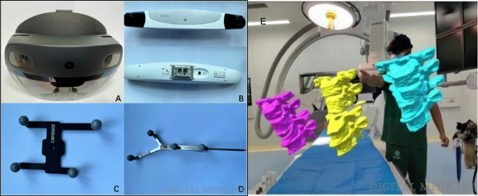
Recent findings: Robotic-assisted pedicle screw placement has been shown to increase the rate of clinically acceptable screw placement while increasing operative time. AI technologies have also shown promise in creating 3D spine imaging while reducing patient radiation exposure. Several models using various imaging modalities have been shown to reliably identify vertebral osteoporotic fractures, stenosis and spine cancers. Complex spinal anatomy and pathology as well as integration of robotics make spine surgery a promising field for the deployment of AI-based imaging technologies. Imaging-based AI projects show potential to enhance diagnostic and surgical efficiency, facilitate trainee learning and improve operative outcomes.
Keywords: Artificial intelligence; Imaging; Oncology; Robotics; Spine surgery; Surgical navigation.
© 2025. The Author(s).
Conflict of interest statement
Declarations. Ethical Approval: This study was determined to be IRB exempt. Informed Consent: Not applicable. Competing Interests: Wellington Hsu is a paid consultant for Asahi, Medtronic Sofamor Danek, and Stryker. He has stock or stock options in Amphix Bio, and is a board or committee member for the Cervical Spine Research Society, Lumbar Spine Research Society and North American Spine Society. All other authors have nothing to disclose.
Figures


References
-
- Pfirrmann CW, et al. Magnetic resonance classification of lumbar intervertebral disc degeneration. Spine (Phila Pa 1976). 2001;26(17):1873–8. - PubMed
-
- Jamaludin A, et al. ISSLS PRIZE IN BIOENGINEERING SCIENCE 2017: automation of reading of radiological features from magnetic resonance images (MRIs) of the lumbar spine without human intervention is comparable with an expert radiologist. Eur Spine J. 2017;26(5):1374–83. - PubMed
-
- Patel NA, et al. Robot-assisted percutaneous pedicle screw placement accuracy compared with alternative guidance in lateral single-position surgery: a systematic review and meta-analysis. J Neurosurg Spine. 2023;39(4):443–51. - PubMed
-
- Fatima N, et al. Safety and accuracy of robot-assisted placement of pedicle screws compared to conventional free-hand technique: a systematic review and meta-analysis. Spine J. 2021;21(2):181–92. - PubMed
Publication types
LinkOut - more resources
Full Text Sources
Research Materials
