. 2025 May;31(5):958-966.
doi: 10.3201/eid3105.250021.

Detection of SARS-CoV-2 Reinfections Using Nucleocapsid Antibody Boosting

Detection of SARS-CoV-2 Reinfections Using Nucleocapsid Antibody Boosting

Eduard Grebe et al. Emerg Infect Dis. 2025 May.

Abstract

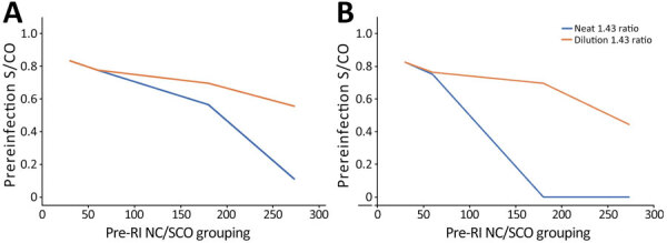
More than 85% of US adults had been infected with SARS-CoV-2 by the end of 2023. Continued serosurveillance of transmission and assessments of correlates of protection require robust detection of reinfections. We developed a serologic method for identifying reinfections in longitudinal blood donor data by assessing nucleocapsid (N) antibody boosting using a total immunoglobulin assay. Receiver operating characteristic curve analysis yielded an optimal ratio of >1.43 (sensitivity 87.1%, specificity 96.0%). When prioritizing specificity, a ratio of >2.33 was optimal (sensitivity 75.3%, specificity 99.3%). In donors with higher anti-N reactivity levels before reinfection, sensitivity was reduced. Sensitivity could be improved by expanding the dynamic range of the assay through dilutional testing, from 38.8% to 66.7% in the highest reactivity group (signal-to-cutoff ratio before reinfection >150). This study demonstrated that longitudinal testing for N antibodies can be used to identify reinfections and estimate total infection incidence in a blood donor cohort.

Keywords: COVID-19; SARS-CoV-2; blood donors; respiratory infections; serology; serosurveillance; severe acute respiratory syndrome coronavirus 2; viruses; zoonoses.


Figures

Figure 1
Figure 1
Individual anti-N S/CO trajectories before and after swab-confirmed reinfection in vaccinated and unvaccinated participants in study of detection of SARS-CoV-2 reinfections using nucleocapsid antibody boosting. A) S/CO trajectories with neat-only anti-N testing of all donors with reinfections in the study. B) Trajectories of test results from 434 donors with reinfections subjected to expanded dynamic range dilutional anti-N testing; neat results only. C) Trajectories of test results from the same 434 donors with reinfections subjected to dilutional anti-N testing; dilutional (expanded dynamic range) testing results only. Images show average anti-N trajectories of donors who experienced reinfections, with and without expanded dynamic range testing, stratified by vaccination status. Time represents days before or after swab-confirmed reinfection (vertical red dashed line). N, nucleocapsid; S/CO, signal-to-cutoff ratio; UU, unvaccinated at the time of first infection and reinfection; UV, unvaccinated at first infection and vaccinated at reinfection; VV, vaccinated at first infection and reinfection.
Figure 2
Figure 2
Effect of prereinfection anti-N S/CO on performance of boosting thresholds in study of detection of SARS-CoV-2 reinfections using nucleocapsid antibody boosting. A) Sensitivity by prereinfection anti-N S/CO using neat and dilutional testing for the unweighted threshold (>1.43); B) sensitivity by prereinfection anti-N S/CO using neat and dilutional testing for the weighted threshold (>2.33). S/CO, signal-to-cutoff ratio.
Figure 3
Figure 3
PPV and NPV in a study of detection of SARS-CoV-2 reinfections using nucleocapsid antibody boosting. A) Unweighted threshold ratio >1.43. B) Weighted threshold ratio >2.33. Predictive values were calculated as a function of percentage of a population of blood donors experiencing SARS-CoV-2 reinfection, at different rates of reinfection. Vertical red lines indicate the proportions reinfected that represent the optimal scenarios for the given threshold ratio, i.e., where PPV and NPV are simultaneously maximized. NPV, negative predictive value; PPV, positive predictive value.

References

    1. Jones JM, Stone M, Sulaeman H, Fink RV, Dave H, Levy ME, et al. Estimated US infection- and vaccine-induced SARS-CoV-2 seroprevalence based on blood donations, July 2020–May 2021. JAMA. 2021;326:1400–9. 10.1001/jama.2021.15161 - DOI - PMC - PubMed
    1. Stone M, Grebe E, Sulaeman H, Di Germanio C, Dave H, Kelly K, et al. Evaluation of commercially available high-throughput SARS-CoV-2 serologic assays for serosurveillance and related applications. Emerg Infect Dis. 2022;28:672–83. 10.3201/eid2803.211885 - DOI - PMC - PubMed
    1. Fink RV, Fisher L, Sulaeman H, Dave H, Levy ME, McCann L, et al. How do we…form and coordinate a national serosurvey of SARS-CoV-2 within the blood collection industry? Transfusion. 2022;62:1321–33. 10.1111/trf.16943 - DOI - PMC - PubMed
    1. Busch MP, Stone M. Serosurveillance for severe acute respiratory syndrome coronavirus 2 (SARS-CoV-2) incidence using global blood donor populations. Clin Infect Dis. 2021;72:254–6. 10.1093/cid/ciaa1116 - DOI - PMC - PubMed
    1. Stone M, Di Germanio C, Wright DJ, Sulaeman H, Dave H, Fink RV, et al.; NHLBI Recipient Epidemiology and Donor Evaluation Study-IV-Pediatric (REDS-IV-P). Use of US blood donors for national serosurveillance of severe acute respiratory syndrome coronavirus 2 antibodies: basis for an expanded national donor serosurveillance program. Clin Infect Dis. 2022;74:871–81. 10.1093/cid/ciab537 - DOI - PMC - PubMed

MeSH terms

Substances