

Case Reports
doi: 10.3201/eid3105.241046.
Tropheryma whipplei Infections, Mexico, 2019-2021
- PMID: 40305382
- PMCID: PMC12044244
- DOI: 10.3201/eid3105.241046
Item in Clipboard
Case Reports
Tropheryma whipplei Infections, Mexico, 2019-2021
Emerg Infect Dis.
2025 May.
Abstract
Whipple's disease is rarely diagnosed in Latin America. We describe 2 patients with Tropheryma whipplei infection diagnosed in Mexico during 2019-2021. Diagnoses were confirmed by histopathology, electron microscopy, immunohistochemistry, and DNA amplification and sequencing analysis of the 16S rRNA gene. Clinicians should be aware of T. whipplei infection and associated syndromes.
Keywords: Mexico; Tropheryma whipplei; Whipple’s disease; bacteria; enteric infections.
Figures

Microscopic and immunohistochemical examination of duodenal tissue samples from a 63-year-old man with Tropheryma whipplei infection, Mexico, 2019. A, B) Hematoxylin and eosin–stained tissue. Microscopic examination showed abundant macrophages in the lamina propria with foamy cytoplasm (A; original magnification ×10); and intracytoplasmic inclusions that stain with PAS (B; original magnification ×40). C) Immunohistochemistry reaction for T. whipplei showed intense positivity in the cytoplasmatic inclusions (original magnification ×40).
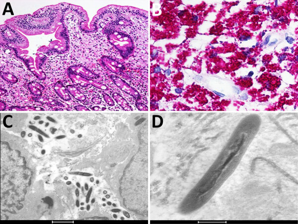
Microscopic and immunohistochemical examination of tissue samples from the ileum in a a 45-year-old man with Tropheryma whipplei infection, Mexico, 2021. A, B) Hematoxylin and eosin–stained ileum tissue. Microscopic examination showed abundant macrophages in the lamina propria with foamy cytoplasm (A; original magnification ×4); and intracytoplasmic inclusions that were intensely PAS-positive (B; original magnification ×40). C, D) Electron microscopy showing rod-shaped T. whipplei in the lamina propria of ileum (C; scale bar = 1 μm), and trilaminar plasma membranes in macrophages (D; scale bar = 200 nm).
References
-
- Whipple GH. A hitherto undescribed disease characterized anatomically by deposits of fat and fatty acids in the intestinal and mesenteric lymphatic tissues. Bull Johns Hopkins Hosp. 1907;18:382–93.
-
- Paddock CD, Fenollar F, Lagier JC, Raoult DA. 21st Century appraisal of Whipple’s disease and Tropheryma whipplei. Clin Microbiol Newsl. 2022;44:123–9. 10.1016/j.clinmicnews.2022.07.001 - DOI
Publication types
MeSH terms
Substances
LinkOut - more resources
Full Text Sources
