

Exosomes from human umbilical cord mesenchymal stem cells promote the growth of human hair dermal papilla cells
- PMID: 40305498
- PMCID: PMC12043141
- DOI: 10.1371/journal.pone.0320154
Exosomes from human umbilical cord mesenchymal stem cells promote the growth of human hair dermal papilla cells
Abstract
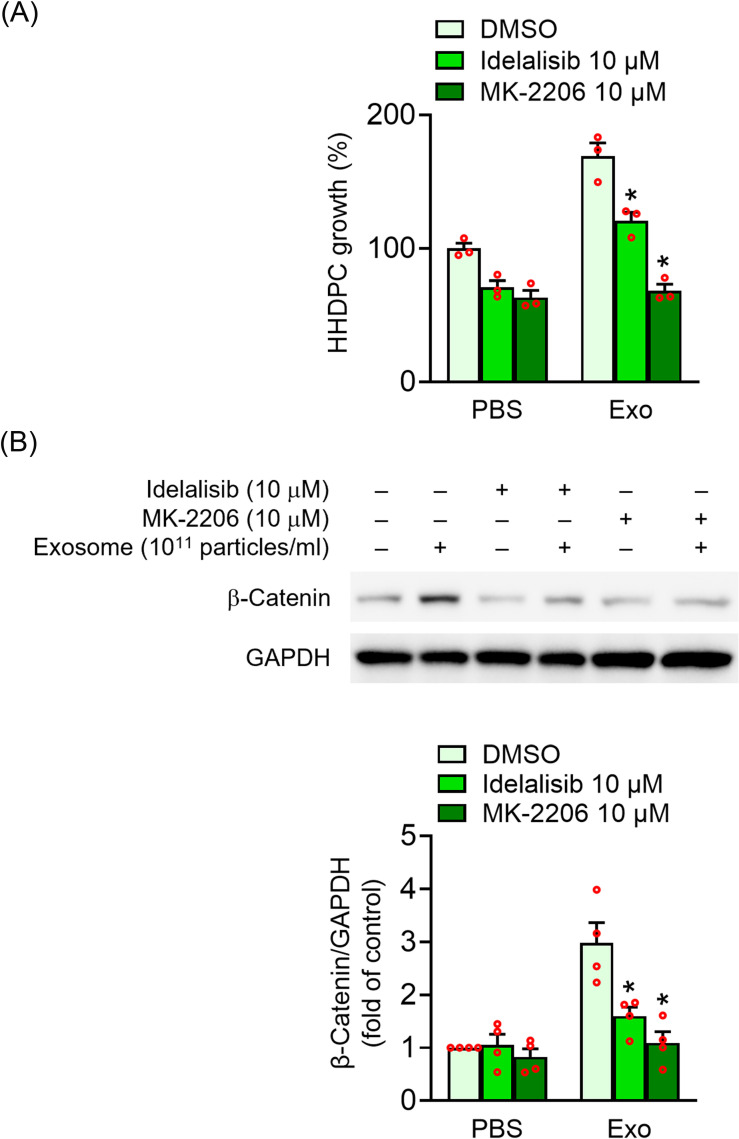
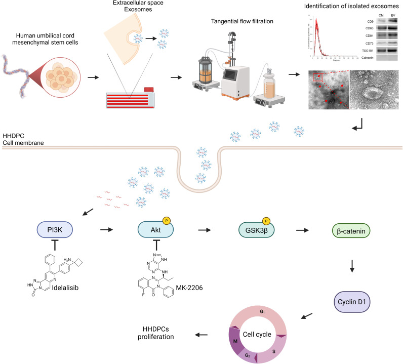
Human hair dermal papilla cells (HHDPCs) play a significant role in hair growth. This study found that human umbilical cord mesenchymal stem cell-derived exosomes (UC-MSC-Es) effectively enhanced cell growth of HHDPCs. UC-MSC-Es has a size range of 30-180 nm and expression of CD9, CD63, CD81, CD73, and TSG101. UC-MSC-Es significantly increased cell populations of HHDPCs in the S and G2/M phases. UC-MSC-Es also increased the expression of cell cycle-related proteins, β-catenin, and cyclin D1. Further mechanistic studies demonstrated that UC-MSC-Es promoted the phosphorylation of Akt and GSK-3β, and the inhibition of PI3K and Akt reduced the proliferative effects of UC-MSC-Es. Collectively, these findings suggest that UC-MSC-Es have a potential effect in treating hair loss through modulating PI3K and Akt-dependent pathways in HHDPCs.
Copyright: © 2025 Chen et al. This is an open access article distributed under the terms of the Creative Commons Attribution License, which permits unrestricted use, distribution, and reproduction in any medium, provided the original author and source are credited.
Conflict of interest statement
This work was funded by ExoOne Bio (SCRPF3M0611). The funders provided exosomes, conducted exosome analysis, and assisted with manuscript preparation. The funders disclose that the research results will be used in hair care products and patent applications. This does not alter our adherence to PLOS ONE policies on sharing data and materials.
Figures


Similar articles
-
Ginsenoside Rg4 Enhances the Inductive Effects of Human Dermal Papilla Spheres on Hair Growth Via the AKT/GSK-3β/β-Catenin Signaling Pathway.J Microbiol Biotechnol. 2021 Jul 28;31(7):933-941. doi: 10.4014/jmb.2101.01032. J Microbiol Biotechnol. 2021. PMID: 34099599 Free PMC article.
-
Suppression of cholangiocarcinoma cell growth by human umbilical cord mesenchymal stem cells: a possible role of Wnt and Akt signaling.PLoS One. 2013 Apr 30;8(4):e62844. doi: 10.1371/journal.pone.0062844. Print 2013. PLoS One. 2013. PMID: 23646150 Free PMC article.
-
PBX homeobox 1 enhances hair follicle mesenchymal stem cell proliferation and reprogramming through activation of the AKT/glycogen synthase kinase signaling pathway and suppression of apoptosis.Stem Cell Res Ther. 2019 Aug 23;10(1):268. doi: 10.1186/s13287-019-1382-y. Stem Cell Res Ther. 2019. PMID: 31443676 Free PMC article.
-
Maintaining Hair Inductivity in Human Dermal Papilla Cells: A Review of Effective Methods.Skin Pharmacol Physiol. 2020;33(5):280-292. doi: 10.1159/000510152. Epub 2020 Oct 14. Skin Pharmacol Physiol. 2020. PMID: 33053562 Review.
-
The therapeutic potential of stem cell-derived exosomes in the ulcerative colitis and colorectal cancer.Stem Cell Res Ther. 2022 Apr 1;13(1):138. doi: 10.1186/s13287-022-02811-5. Stem Cell Res Ther. 2022. PMID: 35365226 Free PMC article. Review.
References
MeSH terms
Substances
LinkOut - more resources
Full Text Sources
Research Materials
Miscellaneous
