. 2025 Apr 30;17(796):eadt9576.
doi: 10.1126/scitranslmed.adt9576. Epub 2025 Apr 30.

Inclusion of a retroviral protease enhances the immunogenicity of VLP-forming mRNA vaccines against HIV-1 or SARS-CoV-2 in mice

Affiliations

Inclusion of a retroviral protease enhances the immunogenicity of VLP-forming mRNA vaccines against HIV-1 or SARS-CoV-2 in mice

Peng Zhang et al. Sci Transl Med. .

Abstract

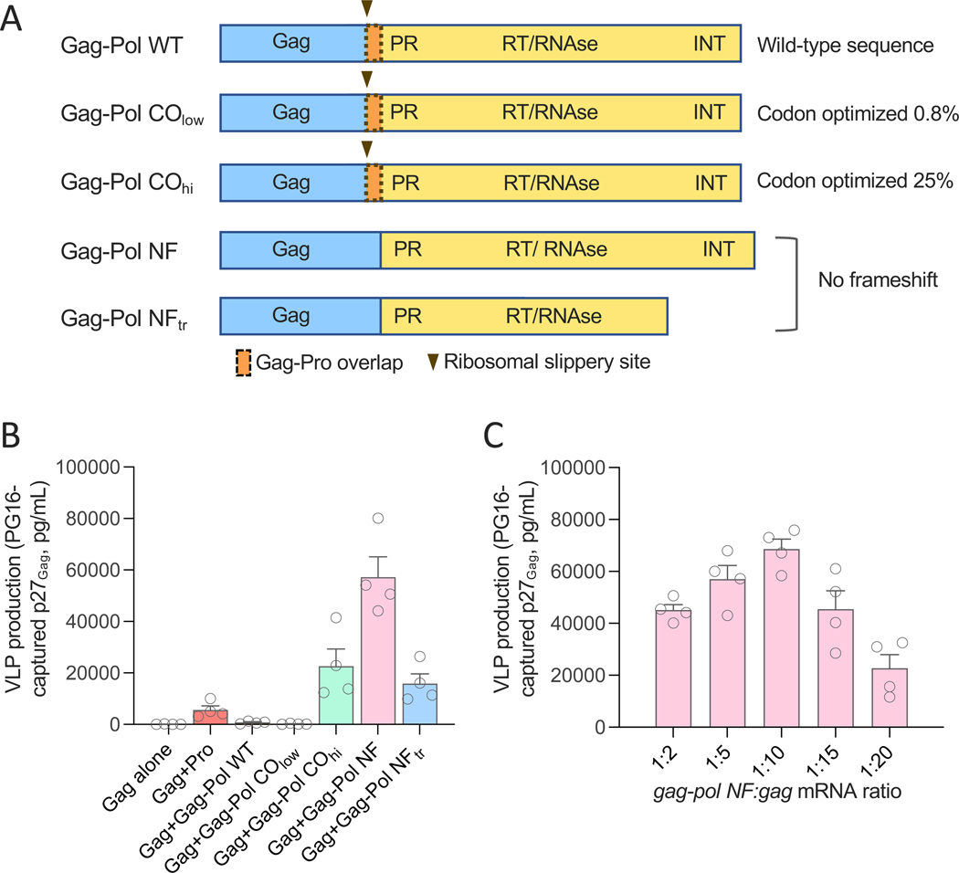
Messenger RNA (mRNA) has emerged as a highly effective and versatile platform for vaccine delivery. We previously designed a virus-like particle (VLP)-forming env-gag mRNA vaccine against human immunodeficiency virus-1 (HIV-1) that elicited envelope-specific neutralizing antibodies and protection from heterologous simian-human immunodeficiency virus (SHIV) infection in rhesus macaques. Here, we introduce a key technological advance to this platform by inclusion of mRNA encoding a retroviral protease to process Gag and produce mature VLPs. Appropriately dosed and timed expression of the protease was achieved using a full-length gag-pol mRNA transcript. Addition of gag-pol mRNA to an HIV-1 env-gag mRNA vaccine resulted in enhanced titers of envelope trimer-binding and neutralizing antibodies in a mouse model. Analogous results were obtained with a hybrid Gag-based, VLP-forming severe acute respiratory syndrome coronavirus 2 (SARS-CoV-2) mRNA vaccine expressing an engineered spike protein. Thus, inclusion of a retroviral protease can increase the immunogenicity of Gag-based, VLP-forming mRNA vaccines against human pathogens.


Conflict of interest statement

Competing interests: P.L., P.Z, S.M.E. and E.N. are inventors on a patent application related to some of the immunogens in this manuscript filed jointly by N.I.H. and Moderna (PCT/US2020/022710, filed on March 13, 2020). S.F., I.R., V.P., E.N., G.S.-J., S.H., and A.C. are employees of Moderna Inc. All other authors declare no competing interests.

Figures

Fig. 1.
Fig. 1.. Addition of mRNA expressing a retroviral protease results in efficient Gag processing but inefficient extracellular VLP production.
(A) Immunoblot analysis of Gag p55 processing in stably Env-expressing HEK293T cells co-transfected with SIV gag mRNA (2 μg per reaction) and pro mRNA at the ratios indicated on the top of the image. The blot was visualized using an anti-SIV Gag antibody that recognizes both uncleaved (p55) and cleaved (p27) SIV Gag. MW, molecular weight. (B) Quantification of total Gag p27 concentration in the culture supernatant of transfected cells. The amount of total Gag p27 (left y axis) was quantified after 48 hours of transfection using an ELISA specific for cleaved p27. Cell viability (right y axis) was measured at 48 hours by flow cytometry using a live-dead dye. (C) Quantification of VLP-associated Gag p27 captured from the culture supernatants of cells transfected as in (B) collected at 48 hours post-transfection using the trimer-specific human bNAb PG16 bound to magnetic beads. The amount of Gag p27 was quantified using the same ELISA as in (B). Data in (B and C) are presented as the mean (± standard error of the mean) from two technical replicates each from two representative experiments.
Fig. 2.
Fig. 2.. Different gag-pol mRNA constructs result in variable expression of the SIV protease and production of mature VLPs.
(A) Schematic representation of five different SIV gag-pol mRNA constructs with or without the ribosomal frameshift mechanism and variable amounts of codon optimization. (B) Quantification of extracellular VLPs produced by stably Env-expressing HEK293T cells co-transfected with SIV gag mRNA (1 μg per reaction) and different gag-pol mRNA constructs (1 μg per reaction) or controls. VLPs were captured from the culture supernatants 48 hours after transfection using the trimer-specific human bNAb PG16 bound to magnetic beads. The amount of VLP-associated Gag p27 was quantified using an ELISA specific for cleaved SIV p27. (C) Quantification of VLP-associated Gag p27 captured from the culture supernatants of cells co-transfected with gag mRNA and decreasing amounts of gag-pol NF mRNA at the indicated gag-pol:gag mRNA ratios; samples were collected at 48 hours post-transfection. Data in (B and C) are presented as the mean (± standard error of the mean) from two technical replicates each from two representative experiments.
Fig. 3.
Fig. 3.. mRNA-expressed immature and mature HIV-1 VLPs display different ultrastructural features.
(A) Cryo-electron tomogram of an immature virus-like particle in which the internal Gag polyprotein lattice is clearly visible and few copies of Env (blue arrows, right column) are displayed on the VLP surface. Serial computational sections through the 3D tomogram are shown from top to bottom. Scale bar, 100 nm. (B) Cryo-electron tomogram of a mature virus-like particle that has undergone proteolytic cleavage of the internal Gag polyprotein. Serial computational sections through the 3D tomogram are shown from top to bottom. Left column, raw tomogram; right column annotated to indicate Env (blue arrows) and Gag core (orange shaded), as well as a second partially formed core (orange arrow). Scale bar, 100 nm.
Fig. 4.
Fig. 4.. Immunogenicity of HIV-1 mRNA vaccines producing immature or mature VLPs in mice.
(A) Design of the immunization study. Each vaccine arm included eight wild-type BALB/c mice. Mice were immunized with in LNP-formulated mRNA encoding HIV-1 426c Env and SIV mac239 Gag with or without Gag-Pol NF. (B) Schematic time course of immunizations (green arrows) and bleedings (red drops). 426c-ΔG3 is deglycosylated at three positions: N276D, N460D and N463D; 426c-ΔG1 at one position: N276D; 426c-WT is fully glycan repaired. (C) Induction of 426c trimer-binding antibodies in the 5 study arms over time was assessed by ELISA. Data are presented as mean values (±SEM) of endpoint titers for each study arm. Statistical comparisons were made by Kruskal-Wallis test followed by post-hoc Dunn’s correction for multiple comparisons with Arm 1. The asterisks indicate significant p-values: * < 0.05; ** < 0.01.
Fig. 5.
Fig. 5.. Neutralizing antibody titers in serum of mice immunized with mRNA producing immature or mature HIV-1 VLPs.
(A) Neutralization of autologous 426c-ΔG3 (left panel) and 426c-ΔG1 (right panel) Env pseudoviruses (PVs) over time in different groups of immunized mice. Data are presented as mean values (±SEM) of IC50 titers for each study arm. (B) Neutralization of HIV-1 426c.ΔG3 (upper row) and 426c.ΔG1 (lower row) PVs by sera from individual immunized mice at day 28, 42, 56, and 126. The data indicate IC50 values shown in box-and-whisker representation with individual points denoted by the circles, upper and lower quartiles by the box, and median values by the horizontal line; whiskers represent the minimum and maximum values. (C) Rate of serum neutralization response against HIV-1 426c.ΔG3 (left panel) and 426c.ΔG1 (right panel) PVs in different study arms. The data indicate the proportion of mice in each group with neutralization titers over background values at each time point. All statistical comparisons were made by non-parametric Kruskal-Wallis test followed by post-hoc Dunn’s correction for multiple comparisons with Arm 1. The asterisks indicate significant p-values: * < 0.05; ** < 0.01; *** <0.001.
Fig. 6.
Fig. 6.. Design of a chimeric SARS-CoV-2 vaccine platform forming mature VLPs.
(A) Schematic representation of the chimeric SARS-CoV-2 spike protein (Spike-S) utilized in this study, encompassing the SIV gp41 cytoplasmic tail, truncated at aa. 745, fused to the C-terminus of the transmembrane domain of the Wuhan-1 spike protein. (B) Quantification of extracellular VLPs produced by HEK293T cells co-transfected with mRNA encoding the chimeric Spike-S protein (1 μg per reaction), SIV gag (1 μg per reaction), and decreasing amounts of Gag-Pol NF (at the ratios indicated on the x axis) collected at 48 hours post-transfection. (C) Quantification of extracellular VLPs produced by HEK293T cells co-transfected with mRNA encoding the chimeric Spike-S protein (1 μg per reaction), SIV gag (1 μg per reaction), and decreasing amounts of Gag-Pol COhi (at the ratios indicated on the x axis) collected at 48 hours post-transfection. VLPs were captured from the culture supernatants 48 hours after transfection using the spike protein-specific monoclonal antibody S118 bound to magnetic beads. The amount of VLP-associated Gag p27 was quantified using an ELISA specific for cleaved SIV p27. Data are presented as the mean (± standard error of the mean) from two technical replicates from a representative experiment out of three performed.
Fig. 7.
Fig. 7.. Immunogenicity of mRNA vaccines producing immature or mature SARS-CoV-2 VLPs in mice.
(A) Design of the immunization study. Each vaccine arm included eight wild-type BALB/c mice. Mice were immunized with in LNP-formulated mRNA encoding chimeric SARS-CoV-2 Spike-S and SIV mac239 Gag with or without Gag-Pol NF. (B) Schematic time course of immunizations (green arrows) and bleedings (red drops). All the immunizations were performed with the same Spike-S mRNA with or without SIV gag and gag-pol mRNA. (C) Induction of SARS-CoV-2 spike trimer-binding antibodies in the 5 study arms over time, as assessed by ELISA. Data are presented as mean values (±SEM) of endpoint titers for each study arm. Statistical comparisons were made by Kruskal-Wallis test followed by post-hoc Dunn’s correction for multiple comparisons with Arm 1. The asterisks indicate significant p-values: * < 0.05; ** < 0.01.
Fig. 8.
Fig. 8.. Neutralizing antibody titers in serum of mice immunized with mRNA producing immature or mature SARS-CoV-2 VLPs.
(A) Neutralization of autologous (Wuhan-1, left panel) and heterologous (B.1.351, right panel) SARS-CoV-2 pseudoviruses (PVs) over time in immunized mice. The data are presented as mean values (±SEM) of half-maximal neutralization titers (IC50) for each study arm. (B) Neutralization of autologous (Wuhan-1, left panel) and heterologous (B.1.351, right panel) PVs by sera from individual immunized mice at day 28, 42, 56, and 126. The data indicate IC50 values shown in box-and-whisker representation with individual points denoted by the circles, upper and lower quartiles by the box and median values by the horizontal line; whiskers represent the minimum and maximum values. (C) Rate of serum neutralization responses with IC50 greater than 1:1000 against SARS-CoV-2 Wuhan-1 (left panel) and B.1.351 (right panel) PVs in different study arms at all time points before the third immunization. The data indicate the proportion of mice in each group with reciprocal neutralization titers over 1000 at days 28, 42, 56, and 85. All statistical comparisons were made by non-parametric Kruskal-Wallis test followed by post-hoc Dunn’s correction for multiple comparisons with Arm 1. The asterisks indicate significant p-values: * < 0.05; ** < 0.01; *** <0.001.

References

    1. Fauci AS, Pantaleo G, Stanley S, Weissman D, Immunopathogenic mechanisms of HIV infection. Ann Intern Med 124, 654–663 (1996). - PubMed
    1. Burton DR, Hangartner L, Broadly Neutralizing Antibodies to HIV and Their Role in Vaccine Design. Annu Rev Immunol 34, 635–659 (2016). - PMC - PubMed
    1. Haynes BF, Burton DR, Mascola JR, Multiple roles for HIV broadly neutralizing antibodies. Sci Transl Med 11, (2019). - PMC - PubMed
    1. Mascola JR, Haynes BF, HIV-1 neutralizing antibodies: understanding nature’s pathways. Immunol Rev 254, 225–244 (2013). - PMC - PubMed
    1. Kwong PD et al. , HIV-1 evades antibody-mediated neutralization through conformational masking of receptor-binding sites. Nature 420, 678–682 (2002). - PubMed

MeSH terms