. 2025 Apr 22;10(11):e190740.
doi: 10.1172/jci.insight.190740. eCollection 2025 Jun 9.

Remdesivir postexposure prophylaxis limits measles-induced "immune amnesia" and measles antibody responses in macaques

Affiliations

Remdesivir postexposure prophylaxis limits measles-induced "immune amnesia" and measles antibody responses in macaques

Andy Kwan Pui Chan et al. JCI Insight. .

Abstract

Measles remains one of the most important causes of worldwide morbidity and mortality in children. Measles virus (MeV) replicates extensively in lymphoid tissue, and most deaths are due to other infectious diseases associated with MeV-induced loss of circulating antibodies to other pathogens. To determine whether remdesivir, a broad-spectrum direct-acting antiviral, affects MeV-induced loss of antibody to other pathogens, we expanded the VirScan technology to detect antibodies to both human and macaque pathogens. We measured the antibody reactivity to MeV and non-MeV viral peptides using plasma from MeV-infected macaques that received remdesivir either as postexposure prophylaxis (PEP) (d3-d14) or as late treatment (LT) (d11-d22) in comparison with macaques that were not treated. Remdesivir PEP, but not LT, limited the loss of antibody to non-MeV pathogens. Remdesivir PEP also limited the antibody response to MeV with a decrease in both the magnitude and breadth of the epitopes recognized. LT had little effect on the magnitude of the MeV-specific antibody response but affected the breadth of the response. Therefore, early, but not late, treatment of measles with the direct-acting antiviral remdesivir prevents the loss of antibody to other pathogens but decreases the response to MeV.

Keywords: Adaptive immunity; Immunology; Infectious disease; Virology.


Figures

Figure 1
Figure 1. Effect of remdesivir administration to MeV-infected rhesus macaques on longitudinal average VARscore and percentage peptide hits (PPH) for human and macaque viral species.
(AD) Longitudinal changes in average VARscores (A and B) and percentage peptide hits (C and D) for selected human (n = 18, 24; A and C) and macaque (n = 6; B and D) viral species were shown in heatmaps. Groups of MeV-infected macaques were not treated (WT MeV; n = 6), treated with RDV either prophylactically (d3–d14; RDV PEP; n = 5), or treated at the onset of disease (d11–d22; RDV LT; n = 3). Sham control animals (n = 4) served as uninfected, untreated controls. Human viral species were selected if the VARscore was over 2 or it PPH was over 10% at any time point in any macaque. The thresholds were chosen based on MeV VARscore and PPH values of pre- and post-MeV infection samples to select for the most important viral species for visualization. All macaque viral pathogens were selected for visualization. DPI, days postinfection; RM, rhesus macaque.
Figure 2
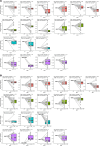
Figure 2. Numeric changes in VARscores from before infection to an early or late time point.
(A and B) Box plots show average change in VARscores of each individual macaque for selected viral species (VARscore >1 at d0) to an early time point (d14/d15) (A) and the last time point (d112/d168/d176) (B). Data for each macaque was represented per box plot. All selected viral species were normalized and, thus, set to have a value of 0 at d0. Each viral species was represented by a gray line. Average numeric change per macaque and the total number of viral species that met the threshold requirement are indicated at the top of each box plot. Color in each box plot represents macaques in the WT MeV (pink), RDV PEP (green), RDV LT (blue), and Sham control (purple) groups. Avg, average; DPI, days postinfection.
Figure 3
Figure 3. Longitudinal changes in VARscores from before infection to all time points.
(A and B) Since d84/d89 sample from 1 RM was not available, the WT MeV group statistics of d0 to d84/d89 contained only 5 animals. Also, 1 RM from the Sham control group was omitted from this analysis, as less than 5 viral species had a VARscore of over 1 at d0. Statistical differences between time points were evaluated by Wilcoxon rank-sum test (A). Nonsignificant results were not shown; *P < 0.05. DPI, days postinfection.
Figure 4
Figure 4. Longitudinal MeV-specific VARscores and average peptide hits per RM group.
(AD) Longitudinal MeV-specific VARscores (A and C) and average peptide hits (B and D) per macaque group. In A and B, gray lines represent values from each macaque, and the red line indicates the average values from all macaques in each group. Wilcoxon rank-sum test was employed to compare values between 2 groups (C and D). Nonsignificant results were not shown; *P < 0.05, **P < 0.01. DPI, days postinfection.
Figure 5
Figure 5. Longitudinal average peptide hits to all MeV proteins and selected viral peptides.
(A and B) Longitudinal average peptide hits to all MeV proteins (A) and selected MeV viral peptides (B) per RM group, visualized by heatmaps. Of note, P encodes 2 nonstructural proteins; V and C, thus P/V/C and P/V, are referring to the same protein peptide tiles of P. In B, MeV viral peptides were selected for analysis when the average hit fold change was > 50 at any time point for any macaque. DPI, days postinfection; hfc, hit fold changes.
Figure 6
Figure 6. Longitudinal average peptide hits to selected MeV peptides in the H and F proteins per RM group.
(A and B) Peptides for H protein (A) and F protein (B) with an average reactivity of HFC > 2 at any time point in any macaque were selected. The top 3 peptide tiles, N477-525, P/V449-504, and H1-56, were reported in Figure 5B and, thus, were excluded from the analysis. Top potential neutralizing targets were highlighted by red boxes. DPI, days postinfection; hfc, hit fold changes.

References

    1. Paules CI, et al. Measles in 2019 - going backward. N Engl J Med. 2019;380(23):2185–2187. doi: 10.1056/NEJMp1905099. - DOI - PubMed
    1. Moss WJ, Griffin DE. What’s going on with measles? J Virol. 2024;98(8):e0075824. doi: 10.1128/jvi.00758-24. - DOI - PMC - PubMed
    1. Moss WJ. Measles. Lancet. 2017;390(10111):2490–2502. doi: 10.1016/S0140-6736(17)31463-0. - DOI - PubMed
    1. Lightwood R, et al. Epithelial giant cells in measles as an acid in diagnosis. J Pediatr. 1970;77(1):59–64. doi: 10.1016/S0022-3476(70)80045-2. - DOI - PubMed
    1. Tatsuo H, et al. SLAM (CDw150) is a cellular receptor for measles virus. Nature. 2000;406(6798):893–897. doi: 10.1038/35022579. - DOI - PubMed

MeSH terms