. 2025 Apr 30;13(4):e009048.
doi: 10.1136/jitc-2024-009048.

CD22 CAR-T cells secreting CD19 T-cell engagers for improved control of B-cell acute lymphoblastic leukemia progression

Affiliations

CD22 CAR-T cells secreting CD19 T-cell engagers for improved control of B-cell acute lymphoblastic leukemia progression

Javier Arroyo-Ródenas et al. J Immunother Cancer. .

Abstract

Background: CD19-directed cancer immunotherapies, based on engineered T cells bearing chimeric antigen receptors (CARs, CAR-T cells) or the systemic administration of bispecific T cell-engaging (TCE) antibodies, have shown impressive clinical responses in relapsed/refractory B-cell acute lymphoblastic leukemia (B-ALL). However, more than half of patients relapse after CAR-T or TCE therapy, with antigen escape or lineage switching accounting for one-third of disease recurrences. To minimize tumor escape, dual-targeting CAR-T cell therapies simultaneously targeting CD19 and CD22 have been developed and validated both preclinically and clinically.

Methods: We have generated the first dual-targeting strategy for B-cell malignancies based on CD22 CAR-T cells secreting an anti-CD19 TCE antibody (CAR-STAb-T) and conducted a comprehensive preclinical characterization comparing its therapeutic potential in B-ALL with that of previously validated dual-targeting CD19/CD22 tandem CAR cells (TanCAR-T cells) and co-administration of two single-targeting CD19 and CD22 CAR-T cells (pooled CAR-T cells).

Results: We demonstrate that CAR-STAb-T cells efficiently redirect bystander T cells, resulting in higher cytotoxicity of B-ALL cells than dual-targeting CAR-T cells at limiting effector:target ratios. Furthermore, when antigen loss was replicated in a heterogeneous B-ALL cell model, CAR-STAb T cells induced more potent and effective cytotoxic responses than dual-targeting CAR-T cells in both short- and long-term co-culture assays, reducing the risk of CD19-positive leukemia escape. In vivo, CAR-STAb-T cells also controlled leukemia progression more efficiently than dual-targeting CAR-T cells in patient-derived xenograft mouse models under T cell-limiting conditions.

Conclusions: CD22 CAR-T cells secreting CD19 T-cell engagers show an enhanced control of B-ALL progression compared with CD19/CD22 dual CAR-based therapies, supporting their potential for clinical testing.

Keywords: Adoptive cell therapy - ACT; Bispecific T cell engager - BiTE; Chimeric antigen receptor - CAR; Hematologic Malignancies; Immunotherapy.


Conflict of interest statement

Competing interests: LA-V is a cofounder of Leadartis, a spin-off company focused on unrelated interests. LA-V and BB are cofounders of STAb Therapeutics, a spin-off company from the Research Institute Hospital 12 de Octubre (imas12). PM is cofounder of OneChain Immunotherapeutics, a spin-off company from the Josep Carreras Leukemia Research Institute.

Figures
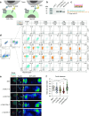
Figure 1
Figure 1. Comparative in vitro study of engineered STAb-19, CAR-22, TanCAR and CAR-STAb Jurkat (J) cells. (a) Schematic representation of the molecular interactions between targeted antigens and CAR or CAR and TCE in the TanCAR and CAR-STAb strategies, respectively. (b) Western blot analysis of CD19-TCE secretion in supernatants from J-CAR-STAb-T (10 times concentrated) and J-STAb-19T cells (net). Samples were subjected to SDS-PAGE and blotting with anti-His-tag antibody. (c) Schematic representation of direct contact co-culture systems to study the specific activation of J-NT-T, J-STAb-T19, J-CAR-T22, J-TanCAR-T and J-CAR-STAb-T cells against a panel of SEM cells genetically engineered using CRISPR-Cas9 technology to selectively silence the expression of one and/or two of the target antigens. (d) Recruitment and activation of transduced (teal) and non-transduced (orange) Jurkat cells cultured with Raji cells at decreasing J:T ratios. One of three independent experiments measuring CD69 by flow cytometry is shown (n=3). Percentages of activated (CD69pos) Jurkat cells are indicated. (e) Topology of the IS induced by J-NT-T, J-STAb-19T, J-CAR-22T, J-TanCAR-T and J-CAR-STAb-T cells. Representative Jurkat/Raji cell conjugates are shown. Conjugates of Jurkat and SEE-loaded Raji cells are used to show the organization of actin at a canonical IS. F-actin distribution to the IS of a confocal section is displayed in pseudocolor. Merged image of F-actin in red and CMAC in cyan is shown for cell identification purposes. Scale bar corresponds to 5 µm. The surface interface of the interaction (pointed by a white square) obtained by 3D-confocal microscopy is shown for F-actin as pseudocolor. The calibration bar of the pseudocolor is indicated. (f) Graph showing F-actin clearance calculated from 3D reconstructions obtained at the IS. Actin clearance value from each individual dot corresponds to the ratio between the central actin cleared area and the total actin area of the 3D IS reconstruction. Each dot represents the value of individual Jurkat/Raji interactions obtained from n=2 independent experiments. Samples were compared by a one-way analysis of variance with a Tukey’s multiple comparison test. Blina, blinatumomab; CAR, chimeric antigen receptor; CMAC, CellTracker Blue; IS, immunological synapses; SEE, Staphylococcal Enterotoxin E; STAb, in situ secretion of T-cell redirecting bispecific antibodies; TanCAR, tandem CAR; TCEs, T cell-engagers; 3D, three-dimensional.
Figure 2
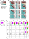
Figure 2. Comparative in vitro cytotoxicity study between CAR-STAb-T and TanCAR-T cells. (a) Transduction efficacy in primary T cells calculated by GFP for CAR-19T, CAR-22T, TanCAR-T and CAR-STAb-T cells or tdTO for STAb-19T cells. Results expressed as mean±SEM of transductions from at least four different healthy donors (n=4). (b, c) Percentages of CD4pos and CD8pos T cells (b) and percentages of CCR7neg CD45RAneg, CCR7neg CD45RApos, CCR7pos CD45RAneg and CCR7pos CD45RApos T cells (c) among NT-T, STAb-19T, CAR-22T, TanCAR-T and CAR-STAb-T cells. Results for (b, c) are mean—SEM of transductions from at least four different healthy donors (n=4). (d) Specific cytotoxicity of non-transduced (NT-T) or transduced (CAR-STAb-T or TanCAR-T) primary T cells against CD19pos/CD22pos (SEMWT), CD19pos/CD22neg (SEM-CD22KO), CD19neg/CD22pos (SEM-CD19KO) or CD19neg/CD22neg (SEM-CD19KO/22KO) target cells at indicated E:T ratio after 48 hours. Data expressed as mean±SEM of one experiment with triplicates (n=3). Cytotoxic activity (e) and IFN-γ secretion determined by ELISA (f) of CAR-STAb-T and TanCAR-T cells co-cultured with NALM6Luc cells under T-cell-limiting conditions (low E:T ratios from 1:1 to 1:512). Data for (e, f) expressed as mean±SEM of two experiments with independent donors in triplicate (n=6). (g) Decreasing numbers of activated effector T (AT) cells (NT-T, CAR-STAb-T or TanCAR-T) were co-cultured with 5×10⁴ NALM6Luc target cells. Increasing numbers of fresh isolated (NA-T) cells from the same donor (bystander T cells) were added to the culture, resulting in the indicated AT:T ratios and maintaining a constant 1:1 E:T ratio. Cytotoxicity (left Y axis) and IFN-γ secretion (right Y axis) are represented. Data expressed as mean±SEM of three experiments with independent donors in triplicate for cytotoxicity curves (n=9) and in duplicate for IFN-γ secretion curves (n=6). (h) Decreasing numbers of activated A-T cells (NT-T, CAR-STAb-T or TanCAR-T) were added to the insert wells of a non-contacting Transwell system at indicated A-T:T ratios, while 5×10⁴ NALM6Luc cells and increasing numbers of NA-T cells were plated in the bottom wells to maintain a constant 1:1 E:T ratio. Data are mean±SEM from two experiments with independent donors, performed in triplicate for cytotoxicity curves (n=6) and duplicate for IFN-γ curves (n=4). The percentage of specific cytotoxicity was calculated after 48 hours of culture by addition of D-luciferin to detect bioluminescence (d–h) and IFN-γ secretion was determined by ELISA (f–h). (i–k) Supernatants from the bystander experiment in (g) were tested in a multiplex bead-based immunoassay for the secretion of granzyme B (i), tumor necrosis factor-α (j) and IL-2 (k). Data for (i–k) expressed as mean±SEM of one experiment performed in triplicate (n=3). Statistical significance was calculated by two-way analysis of variance test corrected with a Tukey’s multiple comparisons test. CAR, chimeric antigen receptor; E:T, effector:target; IFN, interferon; STAb, in situ secretion of T-cell redirecting bispecific antibodies; TanCAR, tandem CAR.
Figure 3
Figure 3. Comparative in vivo efficacy between CAR-STAb-T and TanCAR-T cells in tumor models co-expressing CD19 and CD22. (a) Experimental design of in vivo cytotoxicity in NSG mice receiving 1×10⁶ SEMWT cells, followed by intravenous injection of NT-T, CAR-STAb-T or TanCAR-T cells 4 days later. CAR-STAbpos and TanCARpos cells, represented by GFPpos cells, accounted for 8% of the total T cells injected. (b) Radiance quantification (photons s−1 cm−2 sr−1) at the indicated time points. (c) Bioluminescence images showing disease progression from ventral view. (d) Leukemia progression and T-cell expansion measured by flow cytometry in peripheral blood, bone marrow and brain tissue at indicated time points. (e) Cytokine analysis in plasma and cerebrospinal fluid (CSF) samples from NSG mice treated with NT-T, TanCAR-T and CAR-STAb-T cells. IFN-γ, IFN-β, IL-1β, IL-6 and IL-10 were measured in a multiplex bead-based immunoassay. Results from (b,d,e) expressed as mean±SEM of three mice per group (n=3). (f) Experimental design of in vivo cytotoxicity in NSG mice receiving 1×10⁶ primary B-ALL cells, followed by intravenous injection of NT-T, CAR-STAb-T or TanCAR-T cells 3 weeks later. CAR-STAbpos and TanCARpos cells, accounted by GFPpos cells, comprised 8% of the total injected T cells. (g,h) Percentage of leukemic cells measured by flow cytometry in peripheral blood (g) and bone marrow (h) at indicated time points. (i) Percentage of T cells measured by flow cytometry in bone marrow at week 3. Lines in (g) and bars in (h,i) represent mean±SEM of 4 mice for NT-T group and six mice for both CAR-STAb-T and TanCAR-T groups. (b,d,e,g–i) Statistical significance was calculated by two-way analysis of variance test corrected with a Tukey’s test for multiple comparisons. B-ALL, B-cell acute lymphoblastic leukemia; BLI, bioluminescence signal; BM, bone marrow; CAR, chimeric antigen receptor; E:T, effector:target; IFN, interferon; IL, interleukin; i.v., intravenous; NT-T, non-transduced primary T cells; PB, peripheral blood; PDX, patient-derived xenograft; STAb, in situ secretion of T-cell redirecting bispecific antibodies; TanCAR, tandem CAR.
Figure 4
Figure 4. Cytotoxicity and leukemia control in vitro against heterogeneous tumors containing equal numbers of SEMWT, SEM-CD19KO, and SEM-CD22KO cells (33% SEM cell mix). (a) NT-T, TanCAR-T, CAR-STAb-T or a 1:1 pool of CAR-19T and CAR-22T cells were co-cultured with the 33% SEM cell mix at the indicated ratios. (b) The expression of CD2, CD19 and CD22 was analyzed by flow cytometry after 4, 7 and 11 days to assess cytotoxic activity and leukemia escape over time. Data expressed as mean±SEM of two different experiments with independent donors (n=2). (c) CD19 and CD22 phenotype by flow cytometry at day 4 in a mix containing 33% of SEMWT, SEM-CD19KO, and SEM-CD22KO cells after being stained at day 0 with CellTrace dyes (Far Red for SEMWT, Violet for SEM-CD19KO and CFSE for SEM-CD22KO). (d) The stained 33% SEM cell mix was co-cultured with NT-T, TanCAR-T, CAR-STAb-T or a 1:1 pool of CAR-19T and CAR-22T cells at indicated effector:target ratios. After 4 days, CD19 and CD22 expression was determined by flow cytometry. Percentages of CD19neg, CD22neg, double negative and double positive cells are indicated. CAR, chimeric antigen receptor; NT-T, non-transduced primary T cells; STAb, in situ secretion of T-cell redirecting bispecific antibodies; TanCAR, tandem CAR.
Figure 5
Figure 5. Comparative in vivo efficacy and tumor escape in a tumor model with heterogeneous expression of CD19 and CD22. (a) Experimental design of in vivo cytotoxicity in NSG mice receiving 2.5×10⁵ 33% SEM cell mix (SEMWT, SEM-CD19KO, and SEM-CD22KO cells), followed by intravenous injection of NT-T, CAR-STAb-T, TanCAR-T or a 1:1 pool of CAR-19T and CAR-22T cells 4 days later. The percentage of transduced T cells, accounted by GFPpos or tdTOos cells, was equally adjusted for the different therapies and comprised 14% of the total T cells injected. (b) Radiance quantification (photons s−1 cm−2 sr−1) at the indicated time points. A humane endpoint of 2×10⁷ photons s−1 cm−2 sr−1 was established. (c) Bioluminescence images showing disease progression from ventral view. (d) Overall survival curve after 5 weeks of treatment. Mice were sacrificed according to the radiance humane endpoint of 2×10⁷ photons s−1 cm−2 sr−1. (e) Leukemia progression and T-cell expansion measured by flow cytometry in peripheral blood and bone marrow at endpoint. (f) CD19pos and CD22pos leukemia escape measured by flow cytometry in peripheral blood and bone marrow at endpoint. Percentages indicate CD19 or CD22 positive-stained cells gated from all HLA-ABCpos CD3neg cells. Data in (b,e,f) expressed as mean±SEM of 2 mice for the control group, four mice for the NT-T group and 6 mice for the CAR-STAb-T, TanCAR-T and pooled CAR-T groups. Each dot in (e,f) represents an independent mouse (n=2 for control group, n=4 for NT-T group and n=6 for CAR-STAb-T, pooled CAR-T and TanCAR-T groups). Statistical significance between groups was calculated either by two-way analysis of variance test corrected with a Tukey’s test for multiple comparisons (b) or an unpaired Student’s t-test (e,f). BLI, bioluminescence signal; BM, bone marrow; CAR, chimeric antigen receptor; i.v., intravenous; NT-T, non-transduced primary T cells; PB, peripheral blood; STAb, in situ secretion of T-cell redirecting bispecific antibodies; TanCAR, tandem CAR.

References

    1. Westin JR, Kersten MJ, Salles G, et al. Efficacy and safety of CD19-directed CAR-T cell therapies in patients with relapsed/refractory aggressive B-cell lymphomas: Observations from the JULIET, ZUMA-1, and TRANSCEND trials. Am J Hematol. 2021;96:1295–312. doi: 10.1002/ajh.26301. - DOI - PMC - PubMed
    1. Maude SL, Laetsch TW, Buechner J, et al. Tisagenlecleucel in Children and Young Adults with B-Cell Lymphoblastic Leukemia. N Engl J Med . 2018;378:439–48. doi: 10.1056/NEJMoa1709866. - DOI - PMC - PubMed
    1. Munshi NC, Anderson LD, Jr, Shah N, et al. Idecabtagene Vicleucel in Relapsed and Refractory Multiple Myeloma. N Engl J Med . 2021;384:705–16. doi: 10.1056/NEJMoa2024850. - DOI - PubMed
    1. Neelapu SS, Locke FL, Bartlett NL, et al. Axicabtagene Ciloleucel CAR T-Cell Therapy in Refractory Large B-Cell Lymphoma. N Engl J Med . 2017;377:2531–44. doi: 10.1056/NEJMoa1707447. - DOI - PMC - PubMed
    1. Ortíz-Maldonado V, Rives S, Castellà M, et al. CART19-BE-01: A Multicenter Trial of ARI-0001 Cell Therapy in Patients with CD19+ Relapsed/Refractory Malignancies. Mol Ther. 2021;29:636–44. doi: 10.1016/j.ymthe.2020.09.027. - DOI - PMC - PubMed

MeSH terms

LinkOut - more resources