. 2025 Aug;25(8):e2500028.
doi: 10.1002/mabi.202500028. Epub 2025 Apr 30.

Development of a Decellularized Urinary Bladder Matrix and Heparin-Based Cryogel for Promoting Angiogenesis

Affiliations

Development of a Decellularized Urinary Bladder Matrix and Heparin-Based Cryogel for Promoting Angiogenesis

Dayeon Roo et al. Macromol Biosci. 2025 Aug.

Abstract

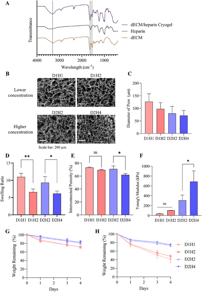
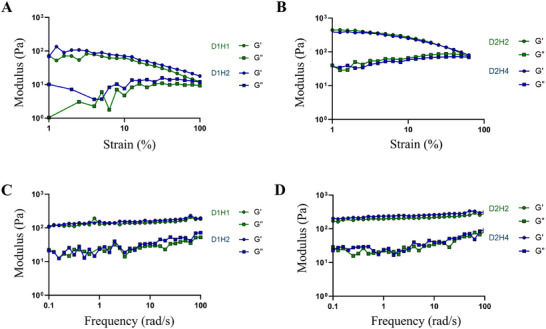
Decellularized extracellular matrix(dECM)-based scaffolds have demonstrated potential in promoting cellular migration and tissue regeneration. In this study, dECM-based cryogel scaffolds are developed with sustained vascular endothelial growth factor (VEGF) release properties to enhance angiogenesis in ischemic tissues. VEGF plays a critical role in angiogenesis by stimulating cell proliferation and migration, but its therapeutic delivery remains challenging due to the need for precise dosing to avoid adverse effects. Cryogels, with their microporous structure, elasticity, and shape-recovery characteristics, offer an ideal platform for controlled VEGF delivery. Using decellularized porcine urinary bladder matrix extracellular matrix (dECM) and heparin, a VEGF-releasing cryogel scaffold is fabricated. The resulting dECM/heparin cryogel is a biocompatible scaffold capable of binding VEGF and releasing it over an extended period. This platform demonstrates significant angiogenic potential both in vitro and in a murine hindlimb ischemia model, highlighting its promise for therapeutic applications in tissue regeneration.

Keywords: cryogel; decellularization; neovascularization; urinary bladder matrix; vascular endothelial growth factor.


Conflict of interest statement

The authors declare no conflict of interest.

Figures

Figure 1
Figure 1
Fabrication of dECM/heparin cryogel. A) Overall schematic illustration of the study. B) Composition of dECM and heparin in each cryogel condition. C) Macroscopic images of dECM/heparin cryogels. The scale bar was 5 mm.
Figure 2
Figure 2
Characterization of dECM/heparin cryogel. A) FT‐IR transmittance analysis of dECM/heparin cross‐linking network. B) Mini‐SEM images showing microstructure of porous cryogels and C) the quantification of dECM/heparin cryogel analyzed by ImageJ. D) Swelling ratio, E) interconnected porosity, and F) Young's modulus of cryogels. Degradation rate at G) 0.2 unit mL−1 of collagenase and H) 1 unit mL−1 of collagenase. The scale bar was 200 µm. * indicated p < 0.05 and ** indicated p < 0.01.
Figure 3
Figure 3
Rheological properties of dECM/heparin cryogel. Storage modulus (G’) and loss modulus (G″) of dECM/heparin cryogels on A,B) strain sweep measured at 1 Hz of frequency and C,D) frequency sweep measured at 5% of strain.
Figure 4
Figure 4
Biocompatibility of dECM/heparin cryogel. A) Cytotoxicity test with LIVE/DEAD assay where live cells were stained in green and dead cells were stained in red. B) Quantification of cell viability. The scale bar was 275 µm. C) Cellular proliferation rate measured with cell counting kit‐8 assay. * indicated p < 0.05, ** indicated p < 0.01, and **** indicated p < 0.0001.
Figure 5
Figure 5
Angiogenic potential in vitro on HUVECs in response to dECM/heparin cryogel loaded with VEGF. A) Representative bright field images of cell scratch assay at 0 and 12 h. B) Quantification of the wound closure analyzed by ImageJ. C) Representative images of tube formation assay stained with calcein‐AM after 12 h. D–G) Quantification of the tube formation assay including D) number of meshes, E) number of nodes, F) number of branches, and G) number of junctions. The scale bar was 650 µm. * indicated p < 0.05, ** indicated p < 0.01, and *** indicated p < 0.001.
Figure 6
Figure 6
Angiogenic potential in a conditioned medium collected from dECM/heparin cryogel loaded with VEGF. A) Cumulative VEGF release rate from dECM/heparin cryogels measured by VEGF ELISA kit. B–E) Analysis on angiogenesis‐related gene expressions of HUVECs with dECM/heparin cryogels loaded with VEGF by RT‐qPCR. * indicated p < 0.05, ** indicated p < 0.01, *** indicated p < 0.001, and **** indicated p < 0.0001.
Figure 7
Figure 7
Angiogenic potential in vivo in an ischemic model. A) Laser Doppler blood perfusion images of ischemic (left) and non‐ischemic (right) hindlimbs on day 0 post‐surgery and on day 28. B) Quantification of blood perfusion by laser Doppler perfusion imaging (LDPI) ratio. C) Necrosis score indicating the salvage score of the hindlimb on day 28. The score ranges from full recovery (score 1) to limb amputation (score 6). D) Immunohistochemistry with α‐smooth muscle cells (α‐SMA) to show blood vessels. E) Quantification of blood vessels. F) Ex vivo H&E staining of a dECM/heparin scaffold with VEGF on day 28. The scale bar was 100 µm. * indicated p < 0.05, ** indicated p < 0.01, and *** indicated p < 0.001.

Similar articles

References

    1. Ryu G. W., Park Y. S., Kim J., Yang Y. S., Ko Y. G., Choi M., JMIR Public Health Surveill 2022, 8, 34908. - PMC - PubMed
    1. Novosel E. C., Kleinhans C., Kluger P. J., Adv. Drug Delivery Rev. 2011, 63, 300. - PubMed
    1. Serini G., Ambrosi D., Giraudo E., Gamba A., Preziosi L., Bussolino F., EMBO J. 2003, 22, 1771. - PMC - PubMed
    1. Bae H., Puranik A. S., Gauvin R., Edalat F., Carrillo‐Conde B., Peppas N. A., Khademhosseini A., Sci. Transl. Med. 2012, 4, 160ps23. - PMC - PubMed
    1. Atienza‐Roca P., Cui X., Hooper G. J., Woodfield T. B. F., Lim K. S., Adv. Exp. Med. Biol. 2018, 1078, 245. - PubMed

MeSH terms

LinkOut - more resources