. 2025 Apr 30;16(1):4068.
doi: 10.1038/s41467-025-59371-4.

O-GlcNAcylation reduces proteome solubility and regulates the formation of biomolecular condensates in human cells

Affiliations

O-GlcNAcylation reduces proteome solubility and regulates the formation of biomolecular condensates in human cells

Senhan Xu et al. Nat Commun. .

Abstract

O-GlcNAcylation plays critical roles in the regulation of protein functions and cellular activities, including protein interactions with other macromolecules. While the formation of biomolecular condensates (or biocondensates) regulated by O-GlcNAcylation in a few individual proteins has been reported, systematic investigation of O-GlcNAcylation on the regulation of biocondensate formation remains to be explored. Here we systematically study the roles of O-GlcNAcylation in regulating protein solubility and its impacts on RNA-protein condensates using mass spectrometry-based chemoproteomics. Unexpectedly, we observe a system-wide decrease in the solubility of proteins modified by O-GlcNAcylation, with glycoproteins involved in focal adhesion and actin binding exhibiting the most significant decrease. Furthermore, O-GlcNAcylation sites located in disordered regions and with fewer acidic and aromatic residues nearby are related to a greater drop in protein solubility. Additionally, we discover that a specific group of O-GlcNAcylation events promotes the dissociation of RNA-protein condensates under heat stress, while some enhance the formation of RNA-protein condensates during the recovery phase. Using site mutagenesis, inhibition of O-GlcNAc transferase, and fluorescence microscopy, we validate that O-GlcNAcylation regulates the formation of biocondensates for YTHDF3 and NUFIP2. This work advances our understanding of the functions of protein O-GlcNAcylation and its roles in the formation of biomolecular condensates.


Conflict of interest statement

Competing interests: The authors declare no competing interests.

Figures

Fig. 1
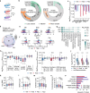
Fig. 1. Experimental procedure to systematically investigate the effect of O-GlcNAcylation on the formation of biocondensates and some experimental results.
a Experimental workflow for the measurement of the solubilities of non-modified and O-GlcNAcylated proteins with or without the RNase treatment. b Quantification of the protein solubilities in the whole proteome. c Overlap between RNase-sensitive proteins found in this work and those by Sridharan et al. d Quantification of the O-GlcNAcylated protein solubilities with or without the RNase treatment. e Percentage of proteins in the whole proteome and O-GlcNAcylated proteins that are soluble/insoluble and RNase-sensitive/insensitive. f Examples of the solubilities of O-GlcNAcylated forms compared with that of the corresponding one in the whole proteome (n = 3 biological replicates). g Examples of the solubilities of the O-GlcNAcylated forms quantified in this work compared with the phosphorylated forms quantified by Sridharan et al. (two-sided Mann-Whitney U tests, n = 3 biological replicates). Ambiguous O-GlcNAcylation site was marked as “amb.” Source data are provided as a Source Data file.
Fig. 2
Fig. 2. Analyses of the RNase-sensitive proteins in cells under heat stress (Heat), during recovery after heat stress (Rec), and the control (Veh).
a Experimental workflow for the preparation of samples under Heat, Rec, and Veh conditions, respectively. b Overall changes of protein solubilities with RNase-treated or mock-treated from Veh to Heat, Heat to Rec, and Veh to Rec. c, Examples of protein solubility change with the RNase treatment in Veh, Heat, and Rec. d Overlap of RNase-sensitive proteins in Veh, Heat, and Rec. e, f Comparing the log2 solubility (e) and the log2 solubility change with the RNase treatment (f) for RNase-sensitive proteins from (d) in different conditions. g Protein clustering results for the RNase-sensitive proteins. RPL subunit stands for ribosomal protein L subunit. h, i Comparison of the log2 solubility change with the RNase treatment for RNA-binding proteins with different functions (h) and for proteins assigned to RNA granule in different tiers by a RNA granule database (i) (n = 3 biological replicates; number of proteins in each category were indicated as N). jn Comparison of the abundance (j), molecular weight (MW, k), GRAVY score (l), isoelectric point (m), and percentage of disordered sequence (n) of RNA-sensitive proteins and other proteins in different conditions (n = 3 biological replicates). o Clustering for the proteins with different percentages in the RNA-protein condensates in the transitions from Veh to Heat and Heat to Rec. Box, 25th/75th percentiles; middle line, median. The differences are assessed using two-sided Mann-Whitney U tests: ∗ P < 0.05, ∗∗ P < 0.01, ∗∗∗ P < 0.001, ∗∗∗∗ P < 0.0001. Whiskers extend to highest/lowest value within 1.5 IQR. Source data are provided as a Source Data file.
Fig. 3
Fig. 3. Effect of O-GlcNAcylation on the protein solubility.
a The effect of O-GlcNAcylation on the protein solubility in Veh, Heat, and Rec. The green dots refer to the O-GlcNAcylated peptides related to a significant solubility decrease, and the orange ones are those with a significant solubility increase. b Comparing the overall effect of O-GlcNAcylation on the protein solubility in Veh, Heat, and Rec (n = 3 biological replicates, two-sided Student’s t-test, B-H adj. P value < 0.05). c Examples of protein solubility changes resulted from O-GlcNAcylation (n = 3 biological replicates). d The effect of O-GlcNAcylation on protein solubility changes in the transitions to different conditions. e Comparison of the O-GlcNAcylation-related solubility changes for proteins with different GO terms (n = 3 biological replicates). f, g Comparison of the O-GlcNAcylation-related solubility change for proteins in RNA-protein granule (f), transcription factors (g) with other glycoproteins. h Evaluation of the O-GlcNAcylation-related solubility change for proteins with various DNA binding domains. Box, 25th/75th percentiles; middle line, median. The differences are assessed using two-sided Mann-Whitney U tests unless mentioned otherwise: ∗ P < 0.05, ∗∗ P < 0.01, ∗∗∗ P < 0.001, ∗∗∗∗ P < 0.0001. Whiskers extend to highest/lowest value within 1.5 IQR. Source data are provided as a Source Data file.
Fig. 4
Fig. 4. Site-specific analysis of the effect of O-GlcNAcylation on protein solubility in cells under heat stress (Heat), during recovery after heat stress (Rec), and the control (Veh).
ad Effect on protein solubility by O-GlcNAcylation with different secondary structures (a), disordered/ordered regions (b), solvent exposed/buried (c), and with or without a protein domain nearby the glycosylation site (d) (n = 3 biological replicates). e, f The occurrence of amino acid residues adjacent to O-GlcNAcylation sites in P1 (e) and P4 (f). g-i, Comparing the average number of acidic (g), aromatic (h), and polar residues (i) near O-GlcNAcylation sites in P1-4. j Distribution of the log2(SolOG/SolPro) difference of the effect of O-GlcNAc on protein solubility in Veh and Heat. k, l Comparing the number of acidic residues adjacent to glycosylation sites (k) and GRAVY score (l) for O-GlcNAc with different effects of protein solubility in Veh and Heat (n = 3 biological replicates). m Distribution of the log2(SolOG/SolPro) difference of the effect of O-GlcNAc on protein solubility in Heat and Rec. n, o, Comparing the number of acidic residues adjacent to glycosylation sites (n) and GRAVY score (o) for O-GlcNAc with different effects of protein solubility in Heat and Rec (n = 3 biological replicates). Box, 25th/75th percentiles; middle line, median (except for (k) and (n), the middle line stands for the mean value). The differences are assessed using two-sided Mann-Whitney U tests: ∗ P < 0.05, ∗∗ P < 0.01, ∗∗∗ P < 0.001, ∗∗∗∗ P < 0.0001. Whiskers extend to the highest/lowest value within 1.5 IQR. Source data are provided as a Source Data file.
Fig. 5
Fig. 5. Investigation of the effect of O-GlcNAcylation on RNA-protein condensates.
ac The effect of O-GlcNAcylation on the formation of condensates with RNA in Veh (a), Heat (b), and Rec (c). d Comparing the effect of O-GlcNAcylation on RNA-protein condensates in Veh, Heat, and Rec (n = 3 biological replicates). e Evaluating the effect of the RNA-protein condensate formation in Veh, Heat, and Rec by the O-GlcNAcylation sites with the top 10% log2(SolOG/SolPro) differences in Heat (n = 3 biological replicates). f, g Evaluating the effect of O-GlcNAcylation sites with the top 10% log2(SolOG/SolPro) differences and other glycosylation sites in Heat on RNA-protein condensates in Veh (f), and on RNA-protein condensates in Rec (g) (n = 3 biological replicates). h Comparison of the effect on RNA-protein condensates in Veh, Heat, and Rec by O-GlcNAcylation sites with the bottom 10% log2(SolOG/SolPro) differences in Rec (n = 3 biological replicates). i, j, Investigation of the effect of O-GlcNAcylation sites with the bottom 10% log2(SolOG/SolPro) differences and other glycosylation sites in Rec on RNA-protein condensates in Veh (i) and Heat (j) (n = 3 biological replicates). k Overlap of O-GlcNAcylated proteins for the glycosylation sites with top 10% log2(SolOG/SolPro) differences in Heat and those with the bottom 10% log2(SolOG/SolPro) differences in Rec. l, m Comparing protein solubility changes by O-GlcNAcylation sites with the top 10% log2(SolOG/SolPro) differences to other glycosites in Heat (l), and the bottom 10% log2(SolOG/SolPro) to other glycosylation sites in Rec (m) (n = 3 biological replicates). n, o, Comparing the number of acidic residues (n) and polar residues (o) nearby the glycosylation sites having top 10% log2(SolOG/SolPro) differences in Heat with other glycosylation sites (n = 3 biological replicates). p Comparing the number of acidic residues adjacent to the glycosylation sites having bottom 10% log2(SolOG/SolPro) differences with other glycosylation sites in Rec (n = 3 biological replicates). Box, 25th/75th percentiles; middle line, median (except for (n), (o), and (p), the middle line stands for the mean value). The differences are assessed using two-sided Mann-Whitney U tests: ∗ P < 0.05, ∗∗ P < 0.01, ∗∗∗ P < 0.001, ∗∗∗∗ P < 0.0001. Whiskers extend to the highest/lowest value within 1.5 IQR. Source data are provided as a Source Data file.
Fig. 6
Fig. 6. Experimental results verified that O-GlcNAcylation regulated the formation of biocondensates for YTHDF3 and NUFIP2.
a Imaging for GFP-tagged YTHDF3 (WT), GFP-tagged YTHDF3 with site mutation (Mutated), and GFP-tagged WT YTHDF3 with the inhibition of OGT (WT with OGTi). Hoechst: the nucleus stained by Hoechst dye; Merged: overlay of the two image channels. Scale bar: 10 µm. b Quantification of the biocondensates in a (n = 5 cells). c Imaging for GFP-tagged NUFIP2 (WT), GFP-tagged NUFIP2 with site mutation (Mutated), and GFP-tagged WT NUFIP2 with the inhibition of OGT (WT with OGTi). d Quantification of the biocondensates in c (n = 5 cells). Center line: mean. The differences are assessed by the two-sided Student’s t-test, and the significance levels are labeled as *** (P < 0.001). Source data are provided as a Source Data file.

Similar articles

Cited by

References

    1. Banani, S. F., Lee, H. O., Hyman, A. A. & Rosen, M. K. Biomolecular condensates: organizers of cellular biochemistry. Nat. Rev. Mol. Cell Biol.18, 285–298 (2017). - PMC - PubMed
    1. Lafontaine, D. L. J., Riback, J. A., Bascetin, R. & Brangwynne, C. P. The nucleolus as a multiphase liquid condensate. Nat. Rev. Mol. Cell Biol.22, 165–182 (2021). - PubMed
    1. Jiang, X. et al. Condensation of pericentrin proteins in human cells illuminates phase separation in centrosome assembly. J. Cell Sci.134, jcs258897 (2021). - PMC - PubMed
    1. Anderson, P., Kedersha, N. & Ivanov, P. Stress granules, P-bodies and cancer. Biochim. Biophys. Acta1849, 861–870 (2015). - PMC - PubMed
    1. Protter, D. S. W. & Parker, R. Principles and properties of stress granules. Trends Cell Biol.26, 668–679 (2016). - PMC - PubMed

LinkOut - more resources