. 2025 Apr 30;15(1):15211.
doi: 10.1038/s41598-025-99795-y.

Effective reduction of unnecessary biopsies through a deep-learning-assisted aggressive prostate cancer detector

Collaborators, Affiliations

Effective reduction of unnecessary biopsies through a deep-learning-assisted aggressive prostate cancer detector

Nuno M Rodrigues et al. Sci Rep. .

Abstract

Despite being one of the most prevalent cancers, prostate cancer (PCa) shows a significantly high survival rate, provided there is timely detection and treatment. Currently, several screening and diagnostic tests are required to be carried out in order to detect PCa. These tests are often invasive, requiring either a biopsy (Gleason score and ISUP) or blood tests (PSA). Computational methods have been shown to help this process, using multiparametric MRI (mpMRI) data to detect PCa, effectively providing value during the diagnosis and monitoring stages. While delineating lesions requires a high degree of experience and expertise from the radiologists, being subject to a high degree of inter-observer variability, often leading to inconsistent readings, these computational models can leverage the information from mpMRI to locate the lesions with a high degree of certainty. By considering as positive samples only those that have an ISUP≥2 we can train aggressive index lesion detection models. The main advantage of this approach is that, by focusing only on aggressive disease, the output of such a model can also be seen as an indication for biopsy, effectively reducing unnecessary biopsy screenings. In this work, we utilize both the highly heterogeneous ProstateNet dataset, and the PI-CAI dataset, to develop accurate aggressive disease detection models.


Figures

Fig. 1
Fig. 1
Connected component analysis. Connected components analysis for both aggressive (ISUP formula image 2) label masks of the ProstateNet and PI-CAI datasets.
Fig. 2
Fig. 2
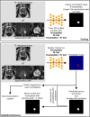
Visualization of the training and validation/inference protocol for the models described in this work. Training was performed using either T2-weighted or biparametric MRI studies belonging to either ProstateNet (PNet), PI-CAI or ProstateNet + PI-CAI (PNetCAI) to detect lesions annotated by radiologists. The validation/inference protocol consists in detecting lesions, extracting the most relevant lesion candidates and considering only lesions with an overlap of at least 10% with the whole prostate gland as inferred by a deep-learning model for prostate segmentation. The patient aggressive lesion probability is then used in a recommendation system, while the binary/probabilistic prediction is used for visualization.
Fig. 3
Fig. 3
Distribution of CAD recommendations, stratified by training and testing dataset. (A) Distribution of annotated (no. of lesions in x-axes) and detected (no. of detected lesions in y-axes) lesions. (B) Relative frequencies of different predictions from the CAD system. For both (A,B) the colors correspond to a classification relating to whether or not this recommendation would lead to a change in the diagnostic algorithm proposed to the patient.
Fig. 4
Fig. 4
Distribution of CAD recommendations, stratified by training for the prospective dataset. (A) Distribution of annotated (no. of lesions in x-axes) and detected (no. of detected lesions in y-axes) lesions. (B) Relative frequencies of different predictions from the CAD system. For both (A,B) the colors correspond to a classification relating to whether or not this recommendation would lead to a change in the diagnostic algorithm proposed to the patient.
Fig. 5
Fig. 5
Effect of lesion size and annotation type on performance for the best performing model (bpMRI). (A) Performance distribution stratified by dataset and lesion size (below or above median). (B) Distribution density for lesion sizes across both datasets. Circles represent the median value while black horizontal lines represent the range between the 1st and 3rd quartiles. (C) Performance distribution stratified by dataset and annotation type (whether the lesion was annotated by a radiologist or by an AI model). (D) Comparison of lesion size with Dice. Each point corresponds to a case, different shapes correspond to different annotation types. Across all plots, golden and blue correspond to PI-CAI and ProstateNet, respectively. p-values in (A,C) correspond to a two-sided Wilcoxon test.
Fig. 6
Fig. 6
Examples of correctly detected and missed cases. (A) Correctly classified and detected lesions. Each row represents a different case selected at random from the correctly detected samples, and the slices shown are those where the index lesion ground truth is most visible in the sequences, (B) missed detected example. The slice choice is the same as the one described previously. For both sets of examples, the ground truth is represented by the white outline, allowing for the view of the target region, and the probability maps are only displayed in the T2W images as to not cover the hyper- and hypo- intense areas of both DWI and ADC sequences.

References

    1. Siegel, R. L., Miller, K. D., Fuchs, H. E. & Jemal, A. Cancer statistics, 2022. CA Cancer J. Clin.72, 7–33 (2022). - PubMed
    1. Resnick, M. J. et al. Long-term functional outcomes after treatment for localized prostate cancer. N. Engl. J. Med.368, 436–445. 10.1056/NEJMoa1209978 (2013). - PMC - PubMed
    1. Scott, R., Misser, S. K., Cioni, D. & Neri, E. PI-RADS v2.1: What has changed and how to report. SA J. Radiol.25, 2062 (2021). - PMC - PubMed
    1. Drost, F.-J. H. et al. Prostate MRI, with or without MRI-targeted biopsy, and systematic biopsy for detecting prostate cancer. Cochrane Database Syst. Rev.4, CD012663 (2019). - PMC - PubMed
    1. Cao, R. et al. Performance of deep learning and genitourinary radiologists in detection of prostate cancer using 3-T multiparametric magnetic resonance imaging. J. Magn. Reson. Imaging54, 474–483 (2021). - PMC - PubMed

Substances