. 2025 Apr 30;163(1):48.
doi: 10.1007/s00418-025-02380-7.

The effects of benzimidazole and electrical stimulation on peripheral nerve regeneration after short- and long-term injury

Affiliations

The effects of benzimidazole and electrical stimulation on peripheral nerve regeneration after short- and long-term injury

Abubaker El Elhaj et al. Histochem Cell Biol. .

Abstract

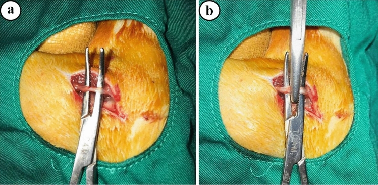
This research investigated the effects of benzimidazole (BZ) and electrical stimulation (ES) on peripheral nerve regeneration after short- and long-term injury and assessed functional recovery by means of stereological, histological, and electrophysiological analyses. Fifty-four male albino Wistar rats were divided into nine groups of six animals each. No treatment or surgery was applied to the control (CONT) group. The sciatic nerve was crushed for 5 s in the short-term injury (STI) and for 60 s in the long-term injury (LTI) groups. In the STI + BZ group and the LTI + BZ group, the rats received 25 mg/kg/day of BZ via oral gavage for 28 days. In the STI + ES and LTI + ES groups, a 3-V current was applied for 20 min daily for 28 days. In the STI + BZ + ES group and the LTI + BZ + ES groups, 3-V ES was applied for 20 min per day for 28 days following oral administration of BZ at 25 mg/kg/day for 28 days. All groups were subjected to electrophysiological, electron microscopic, stereological, and statistical analyses. The stereological analyses revealed a significant increases in the numbers of myelinated axons in the STI + ES groups compared with the STI (p < 0.01). BZ treatment yielded no significant differences in the numbers of myelinated axons in the groups (p > 0.05). Histological evaluation of the STI and LTI groups showed that ES and BZ treatment positively affect the histological structure of the nerve.

Keywords: Benzimidazole; Crush injury; Electrical stimulation; Peripheral nerve; Regeneration; Stereology.


Conflict of interest statement

Declarations. Conflict of interest: The authors declare no competing interests. Ethical approval: This study complied with the tenets of the Declaration of Helsinki and the National Guidelines for Animal Use in Research (Türkiye) and was approved by the Animal Experiments Ethics Committee of Ondokuz Mayıs University under decision no. 2017/10 on 31 March 2017. The experimental procedure was carried out in accordance with the guidelines of the Animal Research Ethics Committee of Ondokuz Mayıs University. The ARRIVE guidelines 2.0 (Animal Research: Reporting of In Vivo Experiments) were strictly followed while doing this experiment.

Figures

Fig. 1
Fig. 1
Schematic representation of the experimental groups
Fig. 2
Fig. 2
a Exposure of the sciatic nerve, b Crush injury induced by clamp
Fig. 3
Fig. 3
a There are notable variations in the sciatic nerve’s mean total number of myelinated axons (*p < 0.05). b Mean axonal area comparison between the groups; no significant differences were seen (p > 0.05). c The mean myelin sheath thickness of nerve fibers in the groups, with a significant difference being observed (*p < 0.05). d A comparison of the mean myelin sheath thickness/axon diameter ratios; no significant differences were observed between the groups (p > 0.05). e There were no significant differences in terms of mean SFI among the groups (p > 0.05). f Differences in mean compound action potential amplitude values, significant differences at the **p < 0.01 and *p < 0.05 levels. g Differences in mean latency; significant differences at the **p < 0.01 level were found between groups
Fig. 4
Fig. 4
Light microscopic images of the sciatic nerves from the study groups. a The myelinated nerve fibers in the CONT group exhibit a normal structure (ax, axon). The diameter of nerve fibers is homogeneous, and some nerve fibers have an impaired myelin sheath. b Most nerve fiber structures in the STI group were severely impaired (dax, degenerated axon). The image shows a few small-sized axons and large spaces between them that are filled by intercellular tissue substances (ct, connective tissue). c Many nerve structures in the STI + ES group were preserved after ES treatment; a thick myelin sheath surrounds the nerve fibers (ax) and healthy structure compared with the injury group. d The majority of nerve fibers in the STI + BZ group were not well protected following BZ administration post-injury, although some fibers had a thick myelin sheath and healthy structure. Much axonal debris (dax) and numerous mast cells (Mc) can be seen. The structure of the vessels (v) appears normal. e Most nerve fiber structures in the STI + ES + BZ group appear normal after injury and treatment with both ES + BZ. They possess a thick myelin sheath and circular spaces in the myelin sheath. f After long-term injury (LTI), most of the nerve structure was filled by connective tissue (ct); it is very difficult to see a cross-section of myelinated nerve fibers. Most of the fibers are very small and difficult to see. g Most nerve structures in the LTI + ES group are profoundly impaired after LTI (dax), but some of them have been partly protected after ES. h After the injury, most nerves in the LTI + BZ group were filled by connective tissue and intercellular tissue substance. Most newly regenerated fibers have a small diameter after injury treated with BZ. i Although most of nerve structures were impaired after injury, many of them were protected by ES + BZ treatment. The formation of a large number of Schwann cells in the injured nerve and a very thin myelin sheath and vessels (v) can be seen in the toluidine blue-stained semi-thin resin section. Scale bars for a, b, c, d, e, f, g, h, and i are 20 µm. Scale bars for the inset of b, c, d, e, g, h, and i are 400 µm, for the inset of f is 200 µm, and for the inset of a is 40 µm
Fig. 5
Fig. 5
Electron microscopic images of sciatic nerves from the study groups. a Images of the sciatic nerves from the CONT group show a well-preserved myelinated (ax) and unmyelinated (arrowhead) nerve fibers. b The structure of myelinated (ax) and unmyelinated (arrowhead) nerve fibers appears normal, and both structures are well preserved. c Images of sciatic nerves treated with ES after injury shows that the nerve fibers’ structure is unaltered. A thick and well-protected myelin sheath can be seen around the fibers, and the general structure of nerve fibers resembles that of the control group. d Following treatment with BZ after injury, the myelin sheath, a thick and well-preserved layer enclosing nerve fiber, appears to be partially normal in structure. e The general structures of nerve fibers treated with ES + BZ after the injury are normal in appearance, and a thick, healthy myelin sheath can be seen around them. f Connective tissue and cells increased after LTI and few number of nerve fibers with thin myelin sheaths are visible, and nerve fibers lost their integrity. g The nerve fibers structure after injury and treatment with ES appears normal. A thick and well-preserved myelin sheath can be seen around small-sized fibers. The borders of each structure in the nerve are clearly visible. h The nerve structure appears partly normal following injury and treatment with BZ, although well-preserved thick myelin sheath can be found around them. However, most of the area in the nerve was filled by connective tissue (ct). i The general nerve structure looks fine, and healthy thick myelin sheath can be found around them. An increased Schwann cell size, especially in their nuclei, can be seen at the bottom center of the image. Scale bar for a, c is 4 µm, for b, f is 2 µm, for d, g, h is 3 µm, and for e is 10 µm. (*) connective tissue
Fig. 6
Fig. 6
Electron microscopic images taken from the sciatic nerves of all LTI types of groups showing macrophage structures in the injury and after treatment. a An image of the only the LTI group is seen; most of the nerve fibers have a considerable distance between each other, and few macrophages are observed. Scale bar 5 µm. b After LTI and exposure to BZ, the size and number of macrophages (ma) are increased. A magnified macrophage (ma) is seen in the inset. The scale bar is 10 µm, and for inset is 3 µm. c, d Applying electric stimulation on the sciatic nerve after LTI creates large-scale macrophages (ma) that engulf debris of nerve fibers in the injured nerve, with sprouting of many new nerves that have intact myelinated nerve fiber structure (ax). The scale bar for c is 6 µm, and for d is 5 µm. e, f Exposing the LTI nerve to ES + BZ combination, macrophages (ma) gain large-scale size, and newly formed nerve fibers reach the intact structure of the myelinated nerve fibers (ax). The scale bar for e is 5 µm, and for f is 3 µm. Ax axon, Ma macrophage

Similar articles

References

    1. Agnew WF, Mccreery DB, Yuen TG, Bullara LA (1999) Evolution and resolution of stimulation-induced axonal injury in peripheral nerve. Muscle Nerve 22:1393–1402. 10.1002/(sici)1097-4598(199910)22:10 - PubMed
    1. Ahlborn P, Schachner M, Irintchev A (2007) One hour of electrical stimulation accelerates functional recovery after femoral nerve repair. Exp Neurol 208:137–144. 10.1016/j.expneurol.2007.08.005 - PubMed
    1. Al-Majed AA, Neumann CM, Brushart TM, Gordon T (2000) Brief electrical stimulation promotes the speed and accuracy of motor axonal regeneration. J Neurosci 20:2602–2608. 10.1523/jneurosci.20-07-02602.2000 - PMC - PubMed
    1. Alvites R, Rita Caseiro A, Santos Pedrosa S, Vieira Branquinho M, Ronchi G, Geuna S, Varejão AS, Colette Maurício A (2018) Peripheral nerve injury and axonotmesis: State of the art and recent advances. Cogent Medicine 5:1466404. 10.1080/2331205X.2018.1466404
    1. Asensio-Pinilla E, Udina E, Jaramillo J, Navarro X (2009) Electrical stimulation combined with exercise increase axonal regeneration after peripheral nerve injury. Exp Neurol 219:258–265. 10.1016/j.expneurol.2009.05.034 - PubMed

LinkOut - more resources