. 2025 May;57(5):964-978.
doi: 10.1038/s12276-025-01443-y. Epub 2025 May 1.

Porphyromonas gingivalis induces Zbp1-mediated macrophages PANoptosis in periodonitis pathophysiology

Affiliations

Porphyromonas gingivalis induces Zbp1-mediated macrophages PANoptosis in periodonitis pathophysiology

Jin Wu et al. Exp Mol Med. 2025 May.

Abstract

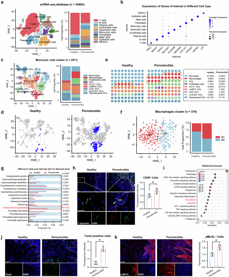
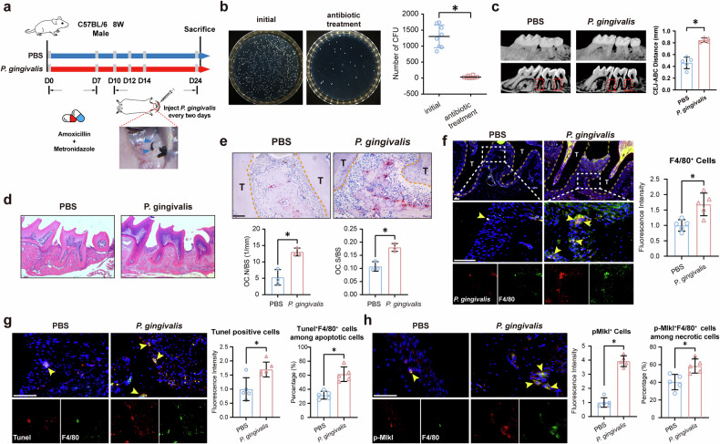
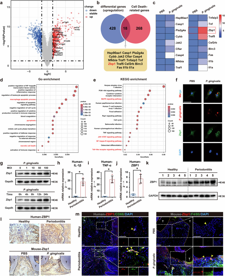
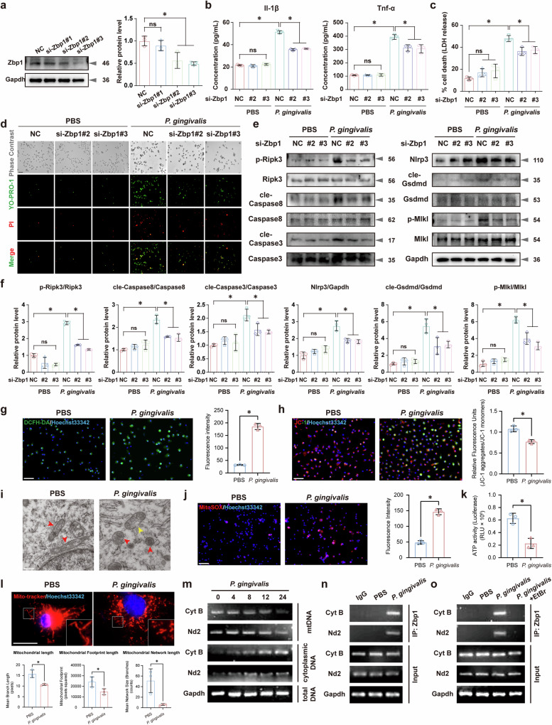
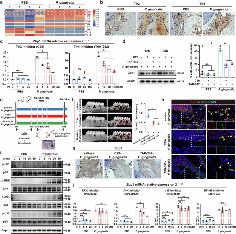
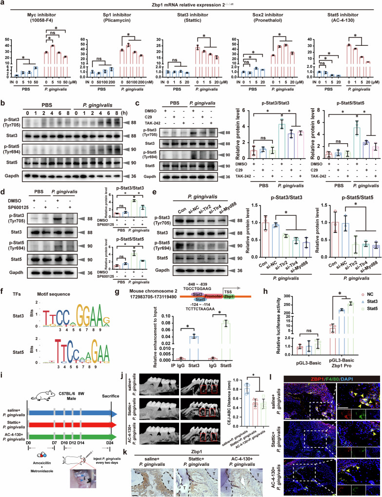
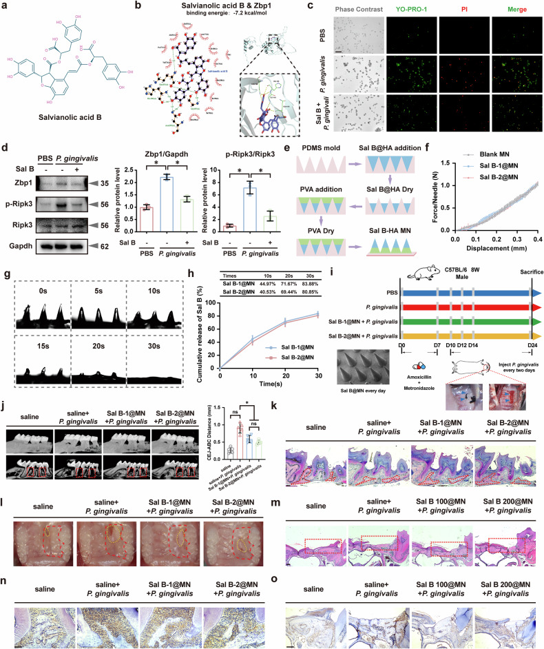
Periodontitis is an oral immunoinflammatory disease, and macrophages play a crucial role in its pathophysiology. However, macrophage death during antibacterial activities will exacerbate inflammation and tissue damage. Porphyromonas gingivalis is a major constituent of subgingival biofilm plaques in periodontitis, but the effects and precise molecular mechanisms by which it triggers macrophage death remain unknown. Here we found that P. gingivalis infection notably activated multiple death pathways in bone-marrow-derived macrophages, including pyroptosis, apoptosis and necrosis. Furthermore, using RNA sequencing, we identified that P. gingivalis infection markedly increased the expression of Z-DNA binding protein 1 (Zbp1) in bone-marrow-derived macrophages. Initially identified as an interferon-induced tumor-associated protein, Zbp1 serves as an upstream sensor that regulates cell death by activating PANoptosis. Mechanistically, P. gingivalis induced a mitochondrial stress response, prompting the release of mitochondrial DNA. This mitochondrial DNA then interacted with Zbp1, consequently augmenting its downstream PANoptosis signals. In addition, P. gingivalis stimulated macrophage Zbp1 expression through the Tlr2/4-JNK-Stat3/5 pathway, exacerbating macrophage death. Importantly, blocking the biosynthesis of endogenous Zbp1 by pharmacological delivery with microneedles improved the survival of P. gingivalis-infected macrophages and inhibited periodontal tissue destruction. These findings highlight Zbp1 as a potential therapeutic target for P. gingivalis-induced periodontitis.


Conflict of interest statement

Competing interests: The authors declare no competing interests.

Figures

Fig. 1
Fig. 1. P. gingivalis infection triggered macrophage infiltration and induced apoptosis and necrosis in periodontal tissues.
a t-Distributed Stochastic Neighbor Embedding (t-SNE) plot and stacked bar plots showing cell subtypes of periodontal samples from the single-cell sequencing dataset GSE171213. b Dot plots showing average expression levels (Z-scaled) and percentage of expressed cells (dot size) of marker genes in major cell types. c Cell subtypes of monocyte clusters. d t-SNE plot representing the macrophage clusters of cells identified across healthy and periodontitis. e Increased abundance of macrophage clusters in periodontitis samples. f Elevated proportion of proinflammatory M1 macrophages in periodontitis. g Top 10 bacterial genera in subgingival plaque. h FISH and IF staining showing increased P. gingivalis accumulation and macrophage infiltration in periodontitis tissues. n = 5. Scale bar, 50 μm. i KEGG enrichment analysis revealing abnormal increase in apoptosis and necrosis pathways in periodontitis macrophages. j, k Elevated TUNEL-positive and p-Mlkl+ cells in periodontitis tissues. n = 5. Scale bar, 50 μm. All data were derived from independent experiments. *P < 0.05.
Fig. 2
Fig. 2. Increased cell apoptosis and necrosis in P. gingivalis-infected mouse periodontal tissues.
a Establishment of a mouse periodontitis model involving the maxillary second molar. n = 5. b Reduced bacterial colonization after antibiotics treatment. n = 10. Representative micro-CT (c) and HE (d) staining images illustrating gingival recession and alveolar bone resorption in P. gingivalis-infected mice. n = 5. Scale bar, 50 μm. e Enhanced TRAP-positive osteoclasts in P. gingivalis-infected periodontitis tissues. n = 5. Scale bar, 50 μm. fh Increased macrophage infiltration (f), cell apoptosis (g) and necrosis (h) in periodontal tissues from mice infected with P. gingivalis. n = 5. Scale bar, 50 μm. All data were derived from independent experiments. *P < 0.05.
Fig. 3
Fig. 3. P. gingivalis infection activated Zbp1 in BMDMs.
a Volcano plot of 1,068 DEGs (500 upregulated, 568 downregulated) in BMDMs treated with PBS or P. gingivalis (n = 4, log2FC ≥1.5 and P < 0.05). b Eighteen upregulated DEGs associated with cell death. c qRT–PCR showing significant upregulation of Zbp1 expression in P. gingivalis-infected BMDMs. n = 3. GO (d) and KEGG (e) enrichment analyses revealing cell death-related pathways abnormalities. f FISH and IF staining showing increased Zbp1 expression in P. gingivalis-infected BMDMs. n = 3. Scale bar, 10 μm. g Higher Zbp1 signals in P. gingivalis-induced BMDMs, displaying both dose- and time-dependent regulation. n = 3. hj qRT–PCR analysis revealing elevated IL-1β, TNF-α and ZBP1 expression in periodontitis tissues. n = 5. *P < 0.05. k Western blotting analyses of Zbp1 expression in gingival tissues from healthy and periodontitis individuals. n = 5. l, m IHC and IF assays confirming upregulated Zbp1 expression in CD68+ and F4/80+ macrophages. n = 5. Scale bar, 50 μm. All data were derived from independent experiments. *P < 0.05.
Fig. 4
Fig. 4. Targeted deletion of Zbp1 in BMDMs protected against PANoptosis induced by P. gingivalis infection.
a Zbp1 knockdown using Zbp1-specific siRNAs. n = 3. ELISA assay showing suppressed IL-1β, TNF-α (b) and LDH (c) release upon inhibition. n = 3. d YO-PRO-1/PI staining showing decreased apoptosis and necrosis with Zbp1 siRNA treatment. n = 3. Scale bar, 50 μm. Western blotting (e) and quantitative analysis (f) demonstrated decreased p-Ripk3, cleaved Caspase8, cleaved Caspase3, Nlrp3, cleaved Gsdmd and p-Mlkl levels after Zbp1 knockdown. n = 3. g Increased ROS levels in BMDMs upon P. gingivalis infection. n = 3. Scale bar, 50 μm. h JC-1 staining showing reduced ΔψM after P. gingivalis infection. n = 3. Scale bar, 50 μm. i Swollen mitochondrial morphology in P. gingivalis-stimulated BMDMs. Scale bar, 500 nm. j, k Elevated mtROS levels and impaired ATP generation in P. gingivalis-infected BMDMs. n = 3. Scale bar, 50 μm. l Fluorescence of MitoTracker revealing increased mitochondrial fragmentation upon P. gingivalis stimulation. n = 3. Scale bar, 10 μm. m PCR assays showing significant cytoplasmic expression of mitochondrial genes Cyt B and Nd2 after P. gingivalis infection. n = 3. n Interactions between mtDNA and Zbp1 identified using a ChIP experiment. n = 3. o Undetectable Cyt B and Nd2 in Zbp1-bound immunoprecipitates after EtBr treatment (10 μg/ml). n = 3. All data were derived from independent experiments. ns, no significance, *P < 0.05.
Fig. 5
Fig. 5. Deletion of Zbp1 protected against PANoptosis activation and periodontal tissue destruction in P. gingivalis-induced mouse periodontitis.
a IHC staining confirming Zbp1 deficiency in periodontal tissues of Zbp1−/− mice. Scale bar, 50 μm. Decreased apoptosis, necrosis (b) and LDH release (c) in BMDMs from Zbp1−/− mice. n = 3. Scale bar, 50 μm. d Establishment of a mouse periodontitis model in WT and Zbp1−/− mice. n = 5. e Representative micro-CT images revealing diminished periodontal tissue destruction in Zbp1−/− mice. n = 5. Diminished periodontal tissue destruction (f) and reduced osteoclastogenesis (g) in Zbp1−/− mice after P. gingivalis challenge. n = 5. Scale bar, 50 μm. h Reduced cleaved Gsdmd, cleaved Caspase3 and p-Mlkl expression in periodontitis lesions of Zbp1−/− mice. Scale bar, 50 μm. i, j Protein analysis from periodontal tissues showing Zbp1 deficiency alleviating upregulation of p-Ripk3, cleaved Caspase8, cleaved Caspase3, Nlrp3, cleaved Gsdmd and p-Mlkl induced by P. gingivalis infection. n = 3. All data were derived from independent experiments. *P < 0.05.
Fig. 6
Fig. 6. P. gingivalis activated Zbp1 in BMDMs via the Tlr2/4–JNK pathways.
a A heat map showing Tlr gene expression profiles in BMDMs stimulated with P. gingivalis. b IHC assays revealing increased levels of Tlr2 and Tlr4 in P. gingivalis-induced periodontal tissues. n = 5. Scale bar, 50 μm. Tlr2 and Tlr4 inhibitors effectively counteracted Zbp1 gene (c) and protein expression (d) in the infected BMDMs. n = 3. e P. gingivalis-infected mouse periodontitis model with daily intraperitoneal administration of C29 (5 mg/kg) and TAK-242 (3 mg/kg). n = 5. Micro-CT (f), IHC (g) and IF staining (h) showing reduced alveolar bone resorption and Zbp1 activation with C29 and TAK-242 treatment. n = 5. Scale bar, 50 μm. i Western blotting showing increased phosphorylation levels of p65, ERK, JNK and p38 in BMDMs after P. gingivalis stimulation. n = 3. j The JNK inhibitor dose-dependently inhibited Zbp1 expression in the infected BMDMs. n = 3. All data were derived from independent experiments. ns, no significance, *P < 0.05.
Fig. 7
Fig. 7. P. gingivalis activated TFs Stat3 and Stat5 to promote Zbp1 transcription.
a P. gingivalis-induced BMDMs showing decreased Zbp1 expression with Stattic and AC-4-130 treatment. n = 3. b Western blotting showing increased phosphorylation of Stat3 and Stat5 after P. gingivalis infection. n = 3. Pharmacological inhibition (c, d) and genetic depletion (e) of the Tlr2/Tlr4–JNK pathway reduced the phosphorylation of Stat3 and Stat5 in P. gingivalis-stimulated BMDMs. n = 3. f Putative binding sites of Stat3 and Stat5 in the Zbp1 promoter regions. g ChIP assays revealing significant Stat3 and Stat5 enrichment in the predicted binding regions of Zbp1 promoter. n = 3. h Luciferase reporter assays validating the regulatory roles of Stat3 and Stat5 in Zbp1 transcription. n = 3. i P. gingivalis-infected mouse periodontitis model with daily intraperitoneal administration of Stattic (5 mg/kg) and AC-4-130 (5 mg/kg). n = 5. Micro-CT images (j), IHC (k) and IF staining (l) showing decreased alveolar bone resorption and Zbp1 activation with Stattic and AC-4-130 treatment. n = 5. Scale bar, 50 μm. All data were derived from independent experiments. ns, no significance, *P < 0.05.
Fig. 8
Fig. 8. MNs delivery of Sal B alleviated P. gingivalis-induced periodontal resorption and promoted mucosal wound healing.
a The chemical structure of Sal B. b Molecular docking simulations revealing Sal B binds favorably to Zbp1. c YO-PRO-1/PI staining showing inhibited apoptosis and necrosis with Sal B intervention. n = 3. Scale bar, 50 μm. d Sal B treatment significantly reduced Zbp1 and p-Ripk3 levels. n = 3. e Encapsulation of Sal B (0.5 mg or 1 mg) into PVA MN patches. f Sal B@MNs demonstrating mechanical strength exceeding 1 N per needle. g Dissolution of the modified MN tips in PBS. h Delivery efficiency of Sal B-1@MN and Sal B-2@MN. n = 3. i P. gingivalis-infected mouse periodontitis and hard palate wound healing models with daily administration of Sal B@MNs. n = 5. Representative images and HE staining illustrating reduced periodontal tissue resorption (j, k) and and near-complete wound healing (l, m) in mice treated with Sal B@MNs. n = 5. Scale bar, 50 μm. IHC images showing lower Zbp1 expression in the periodontitis (n) and hard palate wound healing (o) tissues of Sal B@MNs-treated mice. n = 5. Scale bar, 50 μm. All data were derived from independent experiments. *P < 0.05.

Similar articles

Cited by

References

    1. Slots, J. Periodontitis: facts, fallacies and the future. Periodontol. 200075, 7–23 (2017). - PubMed
    1. Huang, X., Li, Y., Zhang, J. & Feng, Q. Linking periodontitis with inflammatory bowel disease through the oral–gut axis: the potential role of Porphyromonas gingivalis. Biomedicines12, 685 (2024). - PMC - PubMed
    1. Lunar Silva, I. & Cascales, E. Molecular strategies underlying Porphyromonas gingivalis virulence. J. Mol. Biol.433, 166836 (2021). - PubMed
    1. Arafa, E. I. et al. Recruitment and training of alveolar macrophages after pneumococcal pneumonia. JCI Insight7, e150239 (2022). - PMC - PubMed
    1. Galli, G. & Saleh, M. Immunometabolism of macrophages in bacterial infections. Front. Cell Infect. Microbiol.10, 607650 (2020). - PMC - PubMed

MeSH terms

LinkOut - more resources