

Intraocular pressure correlation to progressive retinal nerve fiber layer loss in primary open angle glaucoma as measured by standard and modified goldmann applanation tonometers
- PMID: 40307768
- PMCID: PMC12042340
- DOI: 10.1186/s12886-025-04060-5
Intraocular pressure correlation to progressive retinal nerve fiber layer loss in primary open angle glaucoma as measured by standard and modified goldmann applanation tonometers
Abstract
Purpose: Characterize the relationship between intraocular pressure (IOP) as measured by standard and modified Goldmann prisms and the progressive loss of retinal nerve fiber layer (RNFL) in a cohort of glaucoma patients.
Design: Retrospective cross-sectional cohort data analysis.
Participants: The study included all patients from a database of 1927 eyes, 966 patients with same visit sequential standard and modified Goldmann IOP measurements. From the database, 148 eyes, 75 patients met the inclusion criteria of a diagnosis of primary open angle glaucoma (POAG) with at least 5 sequential quality optical coherence tomographer (OCT) measurements.
Methods: Sequential OCT images were obtained with the spectral domain Zeiss OCT5000. Participants were all diagnosed with POAG by untreated IOP ≥ 22, disk changes, and visual field (HVF) loss consistent with glaucomatous optic neuropathy (GON). Included were 575 Goldmann IOP measurements with standard and modified prisms affixed to the Goldmann applanation tonometer (GAT) armature. A modified prism includes a corneal conforming applanation surface minimizing the cornea's contribution to the IOP measurement. The study included a total of 940 OCT visits with an average of 6.3 visits per eye over an average of 4.9 years. Retinal nerve fiber layer (RNFL) loss rate was calculated by serial linear fit of average RNFL thickness measurements. Demographics as well as central corneal thickness (CCT) and corneal hysteresis (CH) data were also collected.
Outcome measures: Pearson correlation coefficients and random coefficient models were used to evaluate the relationship between mean standard and modified IOP measurements and RNFL thickness measurements over time in POAG subjects. Secondary outcomes of CCT and CH correlation to RNFL were similarly analyzed.
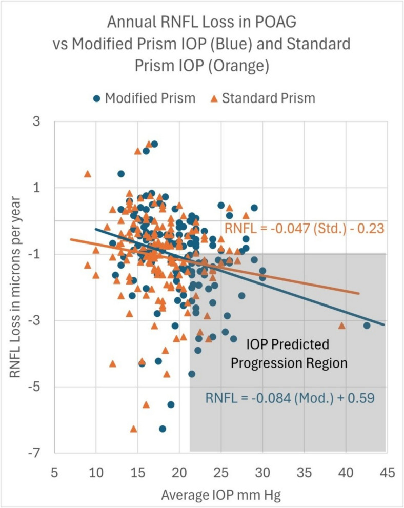
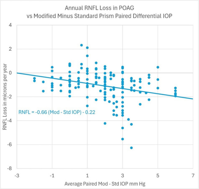
Results: For all 148 POAG eyes, the overall rate of RNFL loss for an average standard GAT IOP of 17.9 mmHg was 1.08 µm per year (p = 0.002). Each 1-mmHg increase in standard GAT IOP was associated with an additional RNFL loss of 0.047 µm per year (r = 0.153, p = 0.06). Each 1-mmHg increase in modified GAT IOP was associated with an additional RNFL loss of 0.084 µm per year (r = 0.289, p = 0.0005). A modified prism IOP measurement ≥ 22 mmHg indicates a 2.57 times greater probability of significant RNFL loss than a standard prism IOP measurement ≥ 22 mmHg, p < 0.0001.
Conclusions: Higher levels of GAT IOP during follow-up were related to higher rates of progressive RNFL loss detected by optic nerve OCT in treated POAG. A modified GAT prism surface demonstrates a significantly increased sensitivity, reliability and differentiation to progressive RNFL loss when compared to a standard GAT prism measured IOP. PRéCIS: A modified applanation surface prism with a corneal conforming shape used on a Goldmann tonometer appears to be a more sensitive and reliable indicator of progressive glaucomatous optic neuropathy as measured by retinal nerve fiber layer changes.
Keywords: Corneal biomechanics; Corneal hysteresis; Glaucoma; Glaucomatous optic neuropathy; OCT; Retinal nerve fiber layer; Tonometry.
© 2025. The Author(s).
Conflict of interest statement
Declarations. Ethics approval and consent to participate: This clinical study was conducted in accordance with the ethical principles contained within Declaration of Helsinki, Protection of Human Volunteers (21 CFR 50), and Obligations of Clinical Investigators (21 CFR 812). The study was completed using retrospective data analysis on deidentified data, therefore IRB approval was not sought. The U.S. Department of Health and Human Services (HHS) regulations under 45 CFR 46.101(b), research involving the collection or study of existing data, documents, records, or specimens can be exempt from IRB review if these sources if the information is recorded by the investigator in such a manner that subjects cannot be identified, directly or through identifiers linked to the subjects. This is outlined in 45 CFR 46.104(d)(4) of the revised Common Rule. Consent for publication: N/A. Competing interests: Only Author Sean McCafferty has a competing interest: The Submitted manuscript titled"Intraocular Pressure Correlation to Progressive Retinal Nerve Fiber Layer Loss in Primary Open Angle Glaucoma as measured by Standard and Modified Goldmann Applanation Tonometers"was completed by referencing studies with funding from an NIH/NEI SBIR grant 1R43 EY026821 - 01. Requirements of this grant are commercialization of potentially beneficial ophthalmic/optometric medical devices/products. The commercialization necessitates intellectual property and a company to produce the product. Commonly in new technology start-up companies (and in this case), an author is also part owner in the intellectual property and the associated company (Intuor Technologies). This is a conflict of interest. However, the authors attest to the stringent efforts made to provide unbiased information provided in this manuscript.
Figures
Similar articles
-
The Relationship between intraocular pressure and progressive retinal nerve fiber layer loss in glaucoma.Ophthalmology. 2009 Jun;116(6):1125-33.e1-3. doi: 10.1016/j.ophtha.2008.12.062. Epub 2009 Apr 19. Ophthalmology. 2009. PMID: 19376584 Free PMC article.
-
The Effect of Age on Increasing Susceptibility to Retinal Nerve Fiber Layer Loss in Glaucoma.Invest Ophthalmol Vis Sci. 2020 Nov 2;61(13):8. doi: 10.1167/iovs.61.13.8. Invest Ophthalmol Vis Sci. 2020. PMID: 33151281 Free PMC article.
-
Impact of Intraocular Pressure Control on Rates of Retinal Nerve Fiber Layer Loss in a Large Clinical Population.Ophthalmology. 2021 Jan;128(1):48-57. doi: 10.1016/j.ophtha.2020.06.027. Epub 2020 Jun 21. Ophthalmology. 2021. PMID: 32579892 Free PMC article.
-
Consensus on Outcome Measures for Glaucoma Effectiveness Trials: Results From a Delphi and Nominal Group Technique Approaches.J Glaucoma. 2016 Jun;25(6):539-46. doi: 10.1097/IJG.0000000000000301. J Glaucoma. 2016. PMID: 26091178
-
Agreement and Reliability of Transpalpebral Tonometers with Goldmann Applanation Tonometer: A Systematic Review and Meta-analysis.Ophthalmol Glaucoma. 2025 May-Jun;8(3):242-256. doi: 10.1016/j.ogla.2024.11.001. Epub 2024 Nov 12. Ophthalmol Glaucoma. 2025. PMID: 39542211
References
-
- Leske M, Heijl A, Hyman L, et al. Predictors of long-term progression in the Early Manifest Glaucoma Trial. Ophthalmology. 2007;114:1965–72. - PubMed
-
- Miglior S, Zeyen T, Pfeiffer N, et al. Results of the European Glaucoma Prevention Study. Ophthalmology. 2005;112:366–75. - PubMed
-
- Kass M, Heuer D, Higginbotham E, et al. The Ocular Hypertension Treatment Study: a randomized trial determines that topical ocular hypotensive medication delays or prevents the onset of primary open-angle glaucoma. Arch Ophthalmol. 2002;120:701–13. - PubMed
-
- The Advanced Glaucoma Intervention Study (AGIS): 7. The relationship between control of intraocular pressure and visual field deterioration. The AGIS Investigators. Am J Ophthalmol 2000;130:429–40. - PubMed
-
- Leske M, Heijl A, Hussein M, et al. Factors for glaucoma progression and the effect of treatment: the Early Manifest Glaucoma Trial. Arch Ophthalmol. 2003;121:48–56. - PubMed
MeSH terms
LinkOut - more resources
Full Text Sources
Medical
Miscellaneous
