. 2025 Apr 30;23(1):326.
doi: 10.1186/s12951-025-03387-x.

Hemoglobin-loaded hollow mesoporous carbon-gold nanocomposites enhance microwave ablation through hypoxia relief

Affiliations

Hemoglobin-loaded hollow mesoporous carbon-gold nanocomposites enhance microwave ablation through hypoxia relief

Yitian Zhang et al. J Nanobiotechnology. .

Abstract

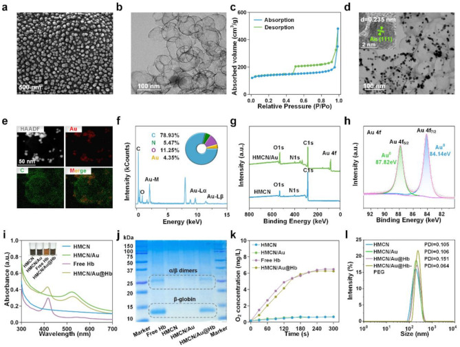
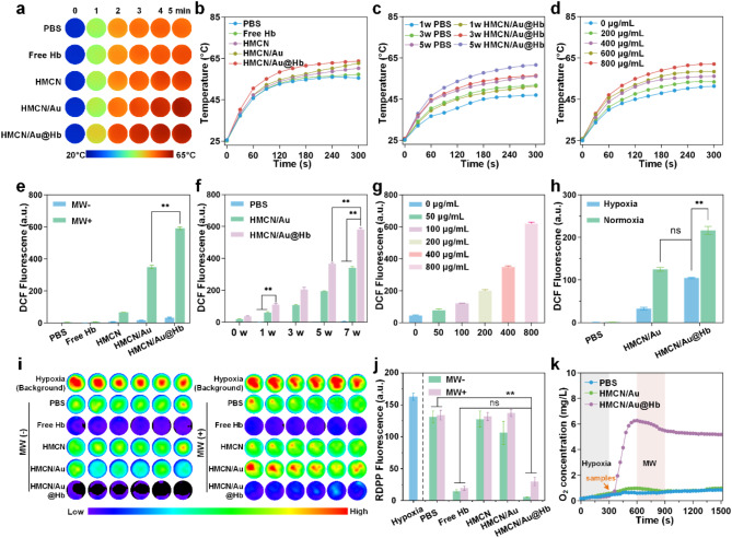
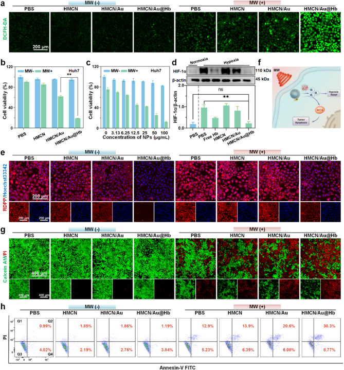
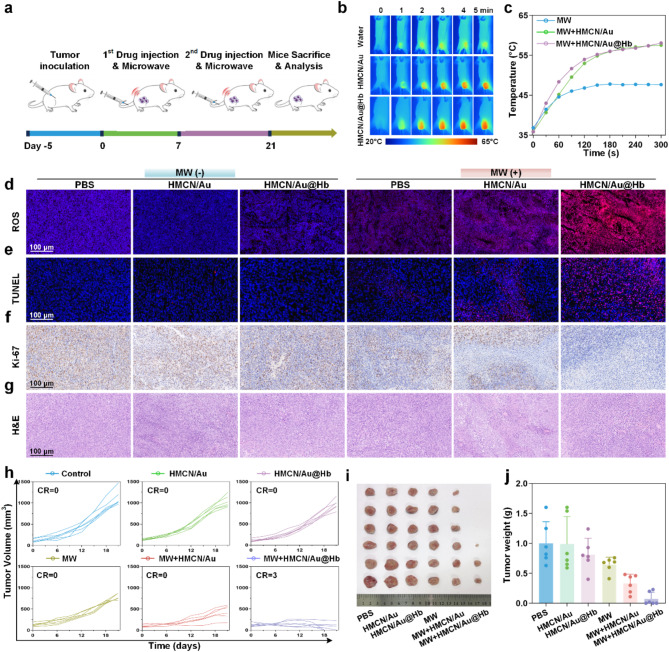
Microwave ablation, as a critical minimally invasive technique for tumor treatment, remains challenging in achieving an optimal balance between incomplete and excessive ablation. In addition to selectively elevating the temperature of tumor lesions through the microwave thermal effect, microwave-responsive nanoparticles can also improve the efficacy of single-session ablation by generating reactive oxygen species (ROS) via the microwave dynamic effect, thereby mitigating the thermal damage to normal tissues caused by high temperature. In this study, ultra-small gold nanoparticles anchored hollow mesoporous carbon nanoparticles (HMCNs) are loaded with hemoglobin (Hb) to serve as microwave ablation nano-sensitizers (HMCN/Au@Hb), which will amplify the microwave dynamic effect by alleviating the hypoxic microenvironment of tumors. Upon microwave irradiation, HMCN/Au@Hb not only improves the microwave-thermal conversion efficiency of tumor lesion but also promotes the ROS generation by increasing oxygen content in the hypoxic tumor microenvironment. More importantly, we found that the hypoxia relief will improve the antitumor response and further enhance the clearance of residual tumor after ablation. Nearly complete ablation was achieved in certain tumor-bearing mice, with no recurrence of the primary tumor observed up to 33 days post-ablation. In comparison to traditional microwave ablation, the survival time of the tumor-bearing mice was significantly extended. Therefore, this work presents an innovative ablation sensitization strategy based on the hypoxia relief and provides a nanosensitizer for microwave ablation integrating great microwave-thermal and dynamic effects along with immune modulation capabilities.

Keywords: Antitumor immune response; Hypoxia relief; Microwave ablation; Microwave dynamic effect; Nanoparticles.


Conflict of interest statement

Declarations. Ethical approval and consent to participate: All the experiments were performed under protocols approved by the Animal Research Ethics Committee of Guangxi university (ethics approval number: GXU-2024-270). Consent for publication: Not applicable. Competing interests: The authors declare no competing interests.

Figures

Scheme 1
Scheme 1
Illustration of synthesis and antitumor mechanism of HMCN/Au@Hb. (a) Synthesis process of HMCN/Au@Hb. Briefly, SiO2 and resorcinol-formaldehyde (RF) oligomers were co-condensed on SiO2 core particles, followed by carbonization under a N2 atmosphere. Subsequently, the silica was etched using NaOH to form hollow mesoporous carbon nanospheres (HMCNs). After anchoring ultra-small gold nanoparticles onto the surface and loading hemoglobin (Hb), the nanocomposites were further functionalized with polyethylene glycol (PEG) to prepare HMCN/Au@Hb. (b) Microwave ablation sensitization employed with HMCN/Au@Hb which integrates great microwave-thermal and dynamic effects along with immune modulation capabilities. HMCN/Au@Hb can effectively absorb microwave energy, leading to the formation of hot spots at the interfaces between HMCN and Au. This phenomenon induces electron transitions and excites trapped oxygen molecules, thereby producing reactive oxygen species (ROS). Meanwhile, HMCN/Au@Hb driven hypoxia relief not only improves ROS production and microwave ablation effect by supplying oxygen, but also synergically enhances the anti-tumor immune effect after ablation by down-regulating the expression of HIF-1α.
Fig. 1
Fig. 1
Preparation and characterization of HMCN/Au@Hb. (a) SEM and (b) TEM images of HMCN. (c) N2 absorption/desorption isotherms of HMCN. (d) TEM images of HMCN/Au (Insert was HRTEM). (e) HAADF-STEM image and elemental mapping for HMCN/Au. (f) Quantitative elemental analysis of HMCN/Au conducted using EDS. The inserts were the comprised element and corresponding atomic frequencies. (g) XPS analysis of the HMCN and HMCN/Au. (h) The high-resolution XPS spectra of Au 4f for HMCN/Au. (i) UV-VIS spectra of free Hb, HMCN, HMCN/Au and HMCN/Au@Hb, respectively (Insert was the color change before and after loading). (j) Coomassie blue staining performed on electrophoresed acrylamide loaded with Hb. (k) Oxygen releasing properties measured by RDPP probe. (l) Size distribution of HMCN, HMCN/Au, HMCN/Au@Hb and PEGylated HMCN/Au@Hb determined by DLS.
Fig. 2
Fig. 2
Microwave responsive and oxygen carrying capacities of HMCN/Au@Hb. (a) Real-time infrared thermal images and (b) corresponding heating profiles of different samples (400 µg/mL) under microwave irradiation (3 W, 5 min). (c) Microwave heating profiles of PBS and HMCN/Au@Hb (400 µg/mL) under microwave irradiation at different power densities for 5 min. (d) Microwave heating profiles of HMCN/Au@Hb at different concentrations under microwave irradiation (3 W, 5 min). (e) ROS detection of different samples (200 µg/mL) irrespective of microwave irradiation (7 W, 5 min). (f) ROS detection of different samples (200 µg/mL) under microwave irradiation (5 min) at different power levels. (g) ROS detection of HMCN/Au@Hb suspensions at different concentrations under microwave irradiation (3 W, 5 min). (h) ROS detection of different samples (PBS, HMCN/Au and HMCN/Au@Hb) at the concentration of 200 µg/mL under hypoxic or normoxic conditions under microwave irradiation (3 W, 5 min). (i) Fluorescence images and (j) quantitative analyses of different samples showing oxygen generation under different conditions. (k) Dynamic oxygen generation of different samples under microwave irradiation (3 W, 5 min). Data were presented as mean values ± SD (n = 3). (* represents p<0.05, ** represents p<0.01)
Fig. 3
Fig. 3
In vitro microwave-induced cytotoxic effect. (a) CLSM images of intracellular ROS level with different treatments (green, fluorescence of DCFH-DA). (b) Cytotoxicity of Huh-7 cells treated with different samples and (c) treated with HMCN/Au@Hb at different concentrations. (d) Western blot demonstrating the HIF-1α expression levels with different treatments. (e) CLSM images of intracellular O2 level with different treatments (blue, fluorescence of Hoechst 33342; red, fluorescence of RDPP; overlay images). (f) Schematic illustration of HMCN/Au@Hb mediated microwave thermal-dynamic effect. (g) Live/Dead co-staining and (h) Annexin V/PI staining of Huh-7 cells after different treatments. Data were presented as mean values ± SD (n = 3). (* represents p<0.05, ** represents p<0.01)
Fig. 4
Fig. 4
In vivo antitumor effect. (a) Schematic illustration of the experimental schedule for the subcutaneous tumor model treatments. (b) Infrared thermal images and (c) the corresponding heating profiles of tumor-bearing mice during microwave irradiation. Histological analysis of sacrificed tumor tissues after a 21-day treatment (d) ROS, (e) TUNEL, (f) Ki-67 and (g) H&E staining. (h) Individual of tumor growth curves in different groups. (i) Photographs (j) and the corresponding weight of tumor tissues at 21st day after treatments. Data were presented as mean values ± SD (n = 6). (* represents p<0.05, ** represents p<0.01)
Fig. 5
Fig. 5
Antitumor immune response. (a) Flow cytometry of DC maturation in TDLNs with indicated treatments. (b) Flow cytometry of T-cell activation in tumors and (c) spleens with indicated treatments. (d) ELISA analysis of the TNF-α, IFN-γ, IL-6, and IL-12p70 levels in serum and tumors. Immunofluorescence images of (e) HIF-1α, (f) Pimonidazole and CD31+, (g) CD8, IFN-γ and Granzyme B exposure in tumors. Data were all presented as mean values ± SD (n = 3). (* represents p<0.05, ** represents p<0.01)

References

    1. Vietti Violi N, Duran R, Guiu B, Cercueil JP, Aube C, Digklia A, Pache I, Deltenre P, Knebel JF, Denys A. Efficacy of microwave ablation versus radiofrequency ablation for the treatment of hepatocellular carcinoma in patients with chronic liver disease: a randomised controlled phase 2 trial. Lancet Gastroenterol Hepatol. 2018;3:317–25. - PubMed
    1. Li L, Zhang X, Zhou J, Zhang L, Xue J, Tao W. Non-Invasive thermal therapy for tissue engineering and regenerative medicine. Small. 2022;18:e2107705. - PubMed
    1. Cao XJ, Wang SR, Che Y, Liu J, Cong ZB, He JF, Wang HL, Liu G, Guo JQ, Hao Y, et al. Efficacy and safety of thermal ablation for treatment of solitary T1N0M0 papillary thyroid carcinoma: A multicenter retrospective study. Radiology. 2021;300:209–16. - PubMed
    1. Zhou H, Fu C, Chen X, Tan L, Yu J, Wu Q, Su L, Huang Z, Cao F, Ren X, et al. Mitochondria-targeted zirconium metal-organic frameworks for enhancing the efficacy of microwave thermal therapy against tumors. Biomater Sci. 2018;6:1535–45. - PubMed
    1. Ma X, Ren X, Guo X, Fu C, Wu Q, Tan L, Li H, Zhang W, Chen X, Zhong H, Meng X. Multifunctional iron-based Metal-Organic framework as biodegradable nanozyme for microwave enhancing dynamic therapy. Biomaterials. 2019;214:119223. - PubMed

LinkOut - more resources