

Monoclonal antibody and B-cell epitope mapping of the VP7 protein in bluetongue virus
- PMID: 40307924
- PMCID: PMC12042368
- DOI: 10.1186/s12985-025-02733-7
Monoclonal antibody and B-cell epitope mapping of the VP7 protein in bluetongue virus
Abstract
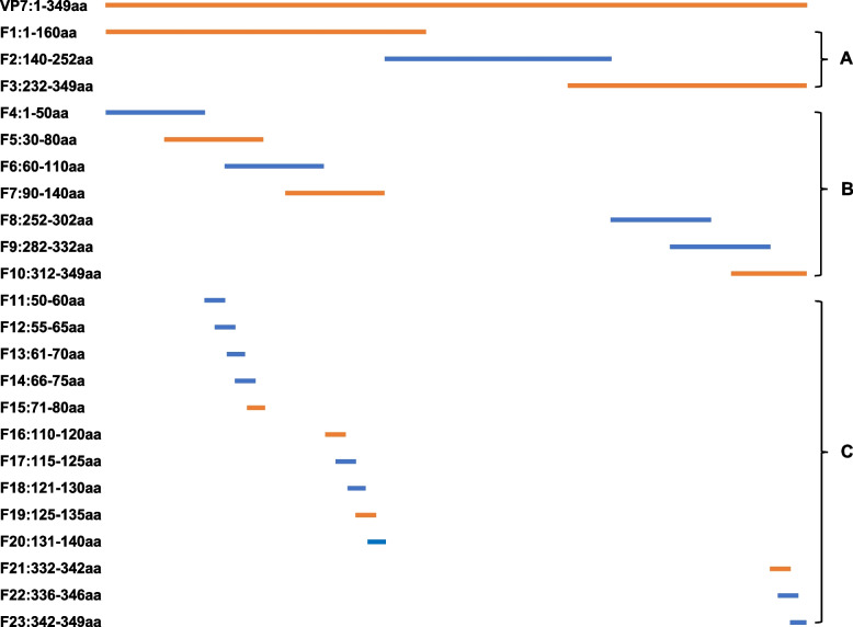
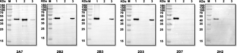
Bluetongue virus (BTV) VP7 is a group-specific protein that is highly conserved in different serotypes. In this study, BALB/c mice were immunized with purified recombinant BTV-1 VP7 protein expressed in E. coli. Then six monoclonal antibodies (mAbs), 2A7, 2B2, 2B3, 2D3, 2D7, and 2H2, against the BTV-1 VP7 protein were produced using hybridoma technology. The reactivity of the mAbs was identified using western blotting, enzyme-linked immunosorbent assay, and immunofluorescence assay. A series of truncated peptides derived from VP7 expressed as glutathione S-transferase fusion proteins were mapped with mAbs by western blotting. The results indicated that 2A7 recognized the epitope 71SAAGINVGPI80, 2B3 recognized 110ARVTGETSTWG120, 2B2 and 2D3 recognized 125PYGFFLETEET135, and 2D7 and 2H2 recognized 332VNPMPGPLTRA342. Amino acid sequence analysis showed that these four epitopes were conserved in 24 typical BTV serotypes. Cross-reaction results showed that mAb 2A7 could recognize the recombinant VP7 protein of BTV-1, African horse sickness virus serotype 1 (AHSV-1), and epidemic hemorrhagic disease virus serotype 1 (EHDV-1). The mAbs 2B2, 2B3, and 2D3 could recognize the recombinant VP7 protein of BTV-1 and EHDV-1, and the mAbs 2D7 and 2H2 specifically recognized the BTV-1 VP7 protein. These specific mAbs and identified B-cell epitopes provided key insights into the structure and function of VP7, while facilitating the development of BTV diagnostics and the design of epitope-based vaccines.
Keywords: B-cell epitope mapping; Bluetongue virus; Monoclonal antibodies; VP7 protein.
© 2025. The Author(s).
Conflict of interest statement
Declarations. Ethics approval and consent to participate: All animals were handled in strict accordance with good animal practice according to the Animal Ethics Procedures and Guidelines of the People’s Republic of China, and the study was approved by The Animal Administration and Ethics Committee of Lanzhou Veterinary Research Institute, Chinese Academy of Agricultural Sciences (Permit No. LVRIAEC-2022–081). Consent for publication: Not applicable. Competing interests: The authors declare no competing interests.
Figures

Similar articles
-
Characterisation of monoclonal antibodies to epizootic hemorrhagic disease virus of deer (EHDV) and bluetongue virus by immunisation of mice with EHDV recombinant VP7 antigen.Res Vet Sci. 1999 Jun;66(3):247-52. doi: 10.1053/rvsc.1998.0282. Res Vet Sci. 1999. PMID: 10333467
-
Analysis of murine B-cell epitopes on bluetongue virus 12 nonstructural protein 1.Appl Microbiol Biotechnol. 2015 Feb;99(3):1309-21. doi: 10.1007/s00253-014-6150-4. Epub 2014 Oct 26. Appl Microbiol Biotechnol. 2015. PMID: 25343975
-
Identification of three novel linear B-cell epitopes on VP7 of African horse sickness virus using monoclonal antibodies.Vet Microbiol. 2024 Nov;298:110258. doi: 10.1016/j.vetmic.2024.110258. Epub 2024 Sep 23. Vet Microbiol. 2024. PMID: 39321671
-
The Global Burden of Emerging and Re-Emerging Orbiviruses in Livestock: An Emphasis on Bluetongue Virus and Epizootic Hemorrhagic Disease Virus.Viruses. 2024 Dec 26;17(1):20. doi: 10.3390/v17010020. Viruses. 2024. PMID: 39861809 Free PMC article. Review.
-
The Genetic Diversification of a Single Bluetongue Virus Strain Using an In Vitro Model of Alternating-Host Transmission.Viruses. 2020 Sep 18;12(9):1038. doi: 10.3390/v12091038. Viruses. 2020. PMID: 32961886 Free PMC article. Review.
References
-
- Carpenter S, Wilson A, Mellor PS. Culicoides and the emergence of bluetongue virus in northern Europe. Trends Microbiol. 2009;17(4):172–8. - PubMed
-
- Belbis G, Zientara S, Bréard E, Sailleau C, Caignard G, Vitour D, et al. Bluetongue virus: from BTV-1 to BTV-27. Adv Virus Res. 2017;99:161–97. - PubMed
-
- Yang H, Gu W, Li Z, Zhang L, Liao D, Song J, et al. Novel putative bluetongue virus serotype 29 isolated from inapparently infected goat in Xinjiang of China. Transbound Emerg Dis. 2021;68(4):2543–55. - PubMed
Publication types
MeSH terms
Substances
Grants and funding
- 22JR5RA024/Science Fund for Creative Research Groups of Gansu Province
- CAAS-ASTIP-2021-LVRI/Innovation Program of Chinese Academy of Agricultural Sciences
- 22CX8NA011/Science Fund for Special Project of Gansu Province
- CARS-37/National Beef Cattle Industrial Technology System
- 2021YFD1800500/National Key Research and Development Program of China
LinkOut - more resources
Full Text Sources
