Case Reports
. 2025 Apr 16:15:1573436.
doi: 10.3389/fonc.2025.1573436. eCollection 2025.

Case Report: Coexistence of an esophageal schwannoma disguised as a leiomyoma with a gastrointestinal stromal tumor of the gastric fundus

Affiliations
Case Reports

Case Report: Coexistence of an esophageal schwannoma disguised as a leiomyoma with a gastrointestinal stromal tumor of the gastric fundus

Yuedong Wang et al. Front Oncol. .

Abstract

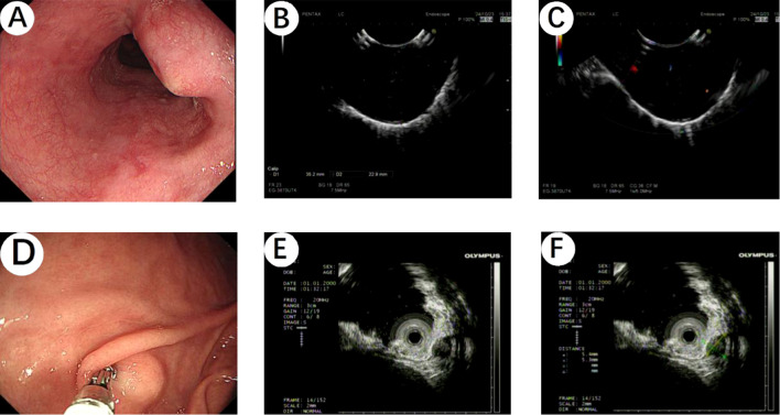
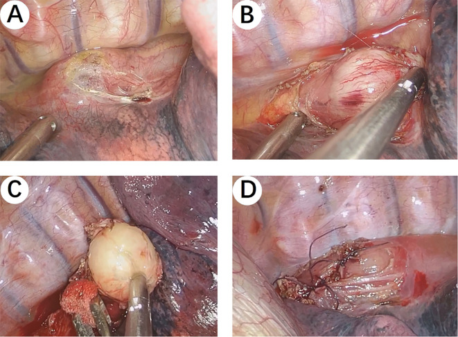
To our knowledge, this is the first reported case of coexisting esophageal schwannoma and gastric fundus gastrointestinal stromal tumor (GIST). This case report describes the diagnostic and treatment process of a patient with esophageal schwannoma who also had a concurrent gastric fundus GIST and was presented to Hebei General Hospital (Hebei, China) in October 2024. The association between the pathogenesis of the two types of submucosal gastrointestinal tumors is unclear, with limited existing evidence in the literature. The esophageal schwannoma was misdiagnosed as a leiomyoma preoperatively, which prompted us to seek new diagnostic modalities to differentiate gastrointestinal submucosal lesions (leiomyomas, GISTs, and schwannomas). Surgical resection is considered the optimal treatment for esophageal schwannoma. The patient underwent a right single-port thoracoscopic esophageal tumor resection and recovered well, subsequently being discharged smoothly from the hospital.

Keywords: case report; differential diagnosis; esophageal schwannoma; gastrointestinal stromal tumor; leiomyoma.


Conflict of interest statement

The authors declare that the research was conducted in the absence of any commercial or financial relationships that could be construed as a potential conflict of interest.

Figures

Figure 1
Figure 1
(A) Chest plain CT scan showing an ovoid soft tissue density shadow (red arrow), approximately 24×21×27mm in size, located at the level of the lower esophagus around the T9 vertebral body. The corresponding lumen at this level showed stenosis. (B, C) Sagittal and coronal reconstruction showing the ovoid soft tissue density shadow (red arrow). (D) The venous phase of the enhanced scan showed that the mass was marked but inhomogeneous enhancement, with a slightly higher enhancement degree than the adjacent esophageal wall. (E, F) Sagittal and coronal reconstruction of the venous phase of the enhanced scan showing the ovoid soft tissue density shadow (red arrow).
Figure 2
Figure 2
(A) Gastroscopy revealed a submucosal mass with a diameter of about 2.0cm at a distance of 30cm from the incisor, with a smooth surface. (B, C) Endoscopic ultrasound showed that the esophageal lesion was located in the muscularis propria, presenting as inhomogeneous hypoechoic, suggesting a leiomyoma. (D) A submucosal mass with a diameter of about 0.6cm was also visible on the greater curvature of the gastric fundus by gastroscopy, with a smooth surface. (E, F) The gastric fundus lesion was located in the muscularis propria, presenting as inhomogeneous hypoechoic, suggesting a stromal tumor.
Figure 3
Figure 3
Intraoperative Images. (A, B) The esophageal lesion was found in the middle and lower part of the thorax, with a hard texture. No invasion of surrounding tissues was observed. (C) The tumor was carefully and completely excised. (D) The esophageal wound was sutured with silk sutures.
Figure 4
Figure 4
Histopathological images. (A) Microscopic image of the esophageal schwannoma taken through a 10x objective on the hematoxylin and eosin-stained histological section shows: a spindle cell tumor. (B) S100 positivity image of the esophageal schwannoma taken through a 100x objective.
Figure 5
Figure 5
Diagnosis and treatment timeline of this case.

Similar articles

References

    1. Jaiswal P, Ijeli AN, Patel D, Jaiswal R, Attar BM, Devani K, et al. . Esophageal schwannoma: A systematic review of 46 cases: 393. Am J Gastroenterol. (2017) 112:S209–10. doi: 10.14309/00000434-201710001-00393 - DOI
    1. Li Y, Teng Y, Wei X, Tian Z, Cao Y, Liu X, et al. . A rare simultaneous coexistence of epithelioid gastrointestinal stromal tumors and schwannoma in the stomach: a case report. Diagn Pathol. (2019) 14:116. doi: 10.1186/s13000-019-0898-x - DOI - PMC - PubMed
    1. Chen S, Zhao Y, Zhao Y. Thoracoscopic resection of a giant esophageal schwannoma: A case report and review of literature. Medicine. (2024) 103:e39507. doi: 10.1097/MD.0000000000039507 - DOI - PMC - PubMed
    1. Matsuki A, Kosugi S, Kanda T, Komukai S, Ohashi M, Umezu H, et al. . Schwannoma of the esophagus: a case exhibiting high 18F-fluorodeoxyglucose uptake in positron emission tomography imaging. Dis Esophagus. (2009) 22:E6–E10. doi: 10.1111/j.1442-2050.2007.00712.x - DOI - PubMed
    1. Park BJ, Carrasquillo J, Bains MS, Flores RM. Giant benign esophageal schwannoma requiring esophagectomy. Ann Thorac Surg. (2006) 82:340–2. doi: 10.1016/j.athoracsur.2005.09.042 - DOI - PubMed

Publication types

LinkOut - more resources