. 2025 Apr 16:12:1564148.
doi: 10.3389/fvets.2025.1564148. eCollection 2025.

Regulatory element map of sheep reproductive tissues: functional annotation of tissue-specific strong active enhancers

Affiliations

Regulatory element map of sheep reproductive tissues: functional annotation of tissue-specific strong active enhancers

Zhu Meng et al. Front Vet Sci. .

Abstract

Introduction: Comprehensive functional annotation of the genome is crucial for elucidating the molecular mechanisms underlying complex traits and diseases. Although functional annotation has been partially completed in sheep, a systematic annotation focused on reproductive tissues remains absent.

Methods: In this study, we integrated 60 transcriptomic and epigenomic datasets from five reproductive tissues. Using a multi-omics approach, we predicted 15 distinct chromatin states and conducted thorough functional annotation.

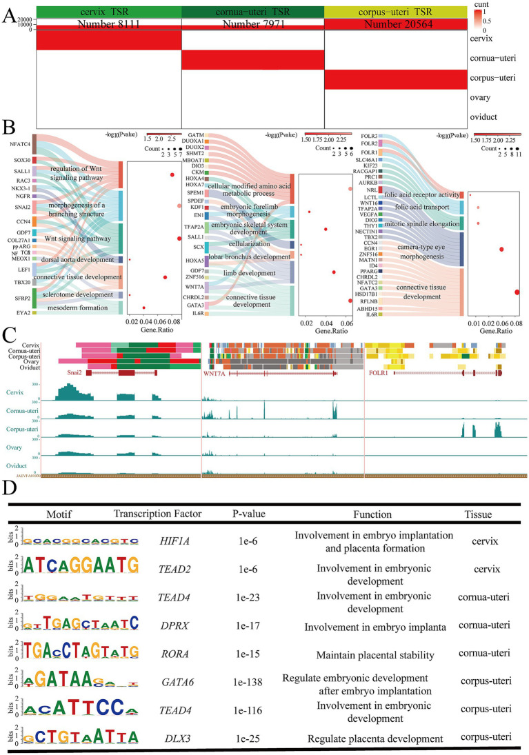
Results: We established the first regulatory element atlas for sheep reproductive tissues and examined the roles of these elements in reproductive traits and disease. In total, we annotated 1,680,172 regulatory elements, including 83,980 tissue-specific strong active enhancers (EnhAs).

Discussion: Enhancers were identified as critical drivers of tissue-specific functions, operating through sequence-specific transcription factor binding and direct regulation of target genes. Key transcription factors associated with reproductive function included INHBA (ovary), KITLG (oviduct), Snai2 (cervix), WNT7A (uterine horn), FOLR1 (uterine body), and SALL1 (shared uterine regions). Additionally, our findings support the potential of sheep as a promising model for investigating embryonic development and miscarriage. This work lays a theoretical foundation for future research into the molecular mechanisms of complex traits and diseases in sheep.

Keywords: enhancer; regulatory element; reproductive tissues; sheep; tissue specific.


Conflict of interest statement

The authors declare that the research was conducted in the absence of any commercial or financial relationships that could be construed as a potential conflict of interest.

Figures

Figure 1
Figure 1
Summary of the data used to construct the epigenomic map of sheep reproductive tissues. (A) Schematic diagram of the tissues and datasets analyzed in this study. The left figure shows a schematic of the reproductive tissue types analyzed, the middle displays the number of epigenomic (ATAC-Seq, H3K4me3, H3K4me1, H3K27me3, and H3K27AC) and transcriptomic (RNA-Seq) samples, and the right figure depicts the main bioinformatics and statistical analyses used in the study. (B) The relationship between tissues based on the Pearson correlation of their normalized signals within a 1 kb window across the entire genome. (C) The average number of peaks for five epigenetic marks across different tissues.
Figure 2
Figure 2
The genome-wide chromatin state landscape across five tissues. (A) Visualization on the UCSC browser (chr1), including chromatin states, CUT&Tag, ATAC-seq, and RNA-seq data. (B) Genome coverage of each chromatin state (proportion, not percentage). (C) The total number of non-redundant regulatory elements (NRRET) and their average size within each chromatin state. (D) The total number of NRRET in five sheep reproductive tissues, across each chromatin state.
Figure 3
Figure 3
Ovarian tissue-specific strong enhancers (EnhA) and their functional annotation. (A) The number and spatial distribution of strong active enhancer (EnhA) in ovarian tissue, along with their enrichment patterns. (B) GO functional enrichment analysis based on the target genes of ovarian EnhAs. (C) Enrichment analysis of transcription factor motifs in ovarian tissue. (D) The chromatin state landscape and mRNA expression of the INHBA (chr4:80,959,294-80,976,837, Ramb_v2.0) locus across five tissues. The vertical scale of the UCSC track represents the normalized RNA-seq signal, ranging from 0 to 210.
Figure 4
Figure 4
Oviducal tissue-specific strong enhancers (EnhA) and their functional annotation. (A) The number and spatial distribution of EnhA in oviducal tissue, along with their enrichment patterns. (B) GO functional enrichment analysis based on the target genes of oviducal EnhAs. (C) Enrichment analysis of transcription factor motifs in oviducal tissue. (D) The chromatin state landscape and mRNA expression of the KITLG (chr3:124,751,722-124,891,391, Ramb_v2.0) locus across five tissues. The vertical scale of the UCSC track represents the normalized RNA-seq signal, ranging from 0 to 150.
Figure 5
Figure 5
Uterine common tissue-specific strong enhancers (EnhA) and functional annotation. (A) The number and spatial distribution of EnhA in uterine common tissue, along with their enrichment patterns. (B) GO functional enrichment analysis based on the target genes of uterine common EnhAs. (C) Enrichment analysis of transcription factor motifs in uterine common tissue. (D) The chromatin state landscape and mRNA expression of the SALL1 (chr14:19,060,302-19,081,400, Ramb_v2.0) locus across five tissues. The vertical scale of the UCSC track represents the normalized RNA-seq signal, ranging from 0 to 50.
Figure 6
Figure 6
Uterus various tissue-specific strong enhancers (EnhA) and their functional annotation. (A) The number and spatial distribution of EnhA in cervix, cornua uteri, and corpus uteri tissue, along with their enrichment patterns. (B) GO functional enrichment analysis based on the target genes of cervix (left figure), cornua uteri (middle figure), and corpus uteri (right figure) EnhAs. (C) Enrichment analysis of transcription factor motifs in cervix (left figure), cornua uteri (middle figure), and corpus uteri (right figure) tissue. (D) The left figure shows the chromatin state landscape and mRNA expression of the SNAI2 (chr9:32,949,607-32,955,054, Ramb_v2.0) locus across five tissues. The vertical scale of the UCSC track represents the normalized RNA-seq signal, ranging from 0 to 300. The middle figure shows the chromatin state landscape and mRNA expression of the WNAT7A (chr19:58,050,035-58,148,599, Ramb_v2.0) locus across five tissues. The vertical scale of the UCSC track represents the normalized RNA-seq signal, ranging from 0 to 10. The right figure shows the chromatin state landscape and mRNA expression of the FOLR1 (chr15:50,521,446-50,538,520, Ramb_v2.0) locus across five tissues. The vertical scale of the UCSC track represents the normalized RNA-seq signal, ranging from 0 to 100.
Figure 7
Figure 7
Human phenotype and mouse phenotype in tissue-specific strong enhancer. (A) Enrichment of specific human phenotypes. (B) Enrichment of specific mouse phenotypes. In the heatmap, the columns represent different tissues, the rows represent different phenotypes, and the color scale indicates the associated tissue for each entry.

References

    1. Mazinani M, Rude B. Population, world production and quality of sheep and goat products. Am J Anim Vet Sci. (2020) 15:291–9. doi: 10.3844/ajavsp.2020.291.299 - DOI
    1. Li Y, Liu Z, Zhang W, Zhu C, Chen X, Zhao Y, et al. . Effects of cloprostenol and oxytocin on ultrasonographic changes of uterine horns in postpartum primiparous Hu sheep. Anim Reprod Sci. (2025) 272:107651. doi: 10.1016/j.anireprosci.2024.107651, PMID: - DOI - PubMed
    1. Gonçalves JD, Dias JH, Machado-Neves M, Vergani GB, Ahmadi B, Pereira Batista RIT, et al. . Transcervical uterine flushing and embryo transfer in sheep: Morphophysiological basis for approaches currently used, major challenges, potential improvements, and new directions (alas, including some old ideas). Reprod Biol. (2024) 24:100920. doi: 10.1016/j.repbio.2024.100920, PMID: - DOI - PubMed
    1. La Y, Tang J, Guo X, Zhang L, Gan S, Zhang X, et al. . Proteomic analysis of sheep uterus reveals its role in prolificacy. J Proteome. (2020) 210:103526. doi: 10.1016/j.jprot.2019.103526, PMID: - DOI - PubMed
    1. Fiorentino G, Cimadomo D, Innocenti F, Soscia D, Vaiarelli A, Ubaldi FM, et al. . Biomechanical forces and signals operating in the ovary during folliculogenesis and their dysregulation: implications for fertility. Hum Reprod Update. (2023) 29:1–23. doi: 10.1093/humupd/dmac031, PMID: - DOI - PubMed

LinkOut - more resources