. 2025 Apr 16:16:1552251.
doi: 10.3389/fphar.2025.1552251. eCollection 2025.

Gui-zhi-fu-ling-wan alleviates bleomycin-induced pulmonary fibrosis through inhibiting epithelial-mesenchymal transition and ferroptosis

Affiliations

Gui-zhi-fu-ling-wan alleviates bleomycin-induced pulmonary fibrosis through inhibiting epithelial-mesenchymal transition and ferroptosis

Zi-Yong Chen et al. Front Pharmacol. .

Abstract

Background: Idiopathic pulmonary fibrosis (IPF) has a higher morbidity and poor prognosis. Gui-Zhi-Fu-Ling-Wan (GFW) is a traditional Chinese herbal formula which exerts anti-inflammatory and anti-oxidative effects. The goal was to determine the protective effect of GFW on bleomycin (BLM)-induced pulmonary fibrosis.

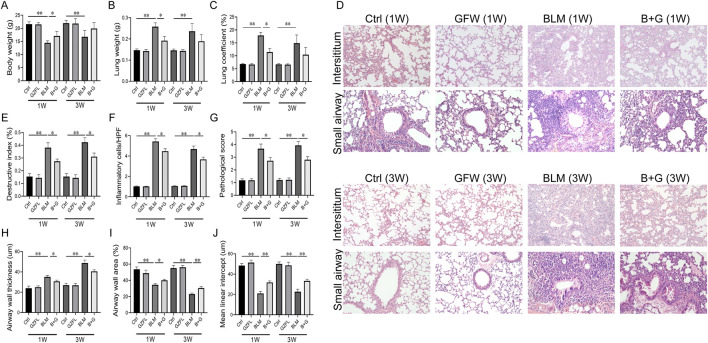
Methods: One hundred and twenty-four mice were randomly divided into eight groups, and orally supplemented with GFW (1 g/kg) in 1 week ago and continuing to 1 week later of single BLM intratracheal injection (5.0 mg/kg). Lung tissues were collected in 7 days and 21 days after BLM injection. BEAS-2B cells were pretreated with GFW (100 μg/mL) for three consecutive days before BLM (10 μg/mL) exposure. Cells were harvested in 12 or 24 h after BLM co-culture.

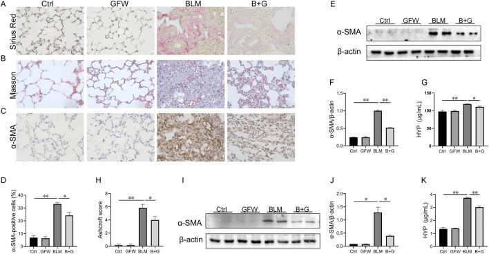
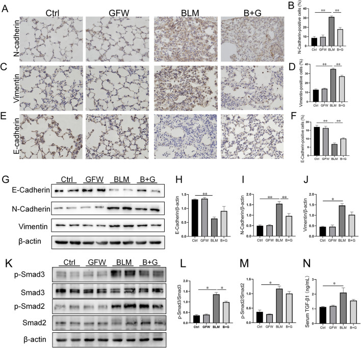
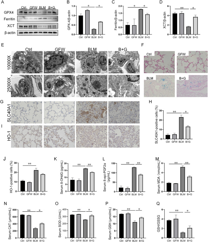
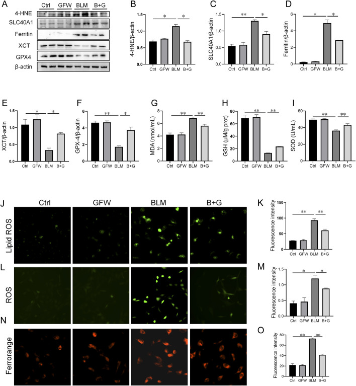
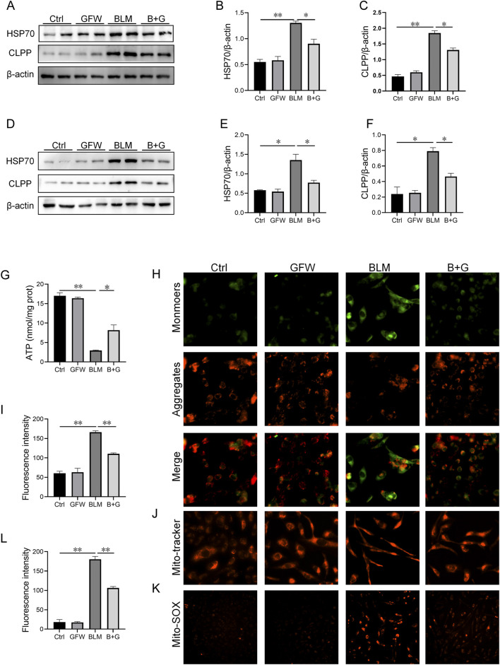
Results: GFW supplementation alleviated BLM-induced alveolar structure destruction and inflammatory cell infiltration in mice lungs. BLM-incurred collagen deposition was attenuated by GFW. In addition, GFW pretreatment repressed BLM-evoked downregulation of E-cadherin, and elevation of N-cadherin and Vimentin in mouse lungs. Besides, BLM-excited GPX4 reduction, ferritin increases, lipid peroxidation, and free iron overload were significantly relieved by GFW pretreatment in mouse lungs and BEAS-2B cells. Notably, BLM-provoked mitochondrial reactive oxygen species (mtROS) excessive production, elevation of mitochondrial stress markers, such as HSP70 and CLPP, and mitochondrial injury, were all abolished in mouse lungs and BEAS-2B cells by GFW pretreatment.

Conclusion: GFW supplementation attenuated BLM-evoked lung injury and pulmonary fibrosis partially through repressing EMT and mtROS-mediated ferroptosis in pulmonary epithelial cells.

Keywords: bleomycin; epithelial-mesenchymal transition; ferroptosis; gui-zhi-fu-ling-wan; mitochondrial reactive oxygen species; pulmonary fibrosis.


Conflict of interest statement

The authors declare that the research was conducted in the absence of any commercial or financial relationships that could be construed as a potential conflict of interest.

Figures

FIGURE 1
FIGURE 1
GFW supplementation attenuated BLM-induced lung injury in mice. Balb/c mice were intratracheally instilled with BLM (5 mg/kg) with or without GFW (1 g/kg) pretreatment. Lung specimens were collected in 7 and 21 days after BLM exposure. The effect of GFW supplementation on BLM-induced lung injury was assessed in mice lungs (A) Body weight (B) Lung weight (C) Lung coefficient (D) Lung interstitium and airway histopathological damage were evaluated by HE staining. Scale bar: 100 µm. Original magnification: ×100 (E) Destructive index (F) The count of inflammatory cells per high-power field (HPF) (G) Pathological score (H) Airway wall thickness (I) Airway wall area (J) Mean linear intercept. All data were expressed as means ± S.E.M. (N = 15). *P < 0.05, **P < 0.01.
FIGURE 2
FIGURE 2
GFW supplementation alleviated BLM-evoked pulmonary fibrosis in mouse lungs. Balb/c mice were intratracheally instilled with BLM (5 mg/kg) with or without GFW (1 g/kg) pretreatment. Lung specimens were collected in 7 and 21 days after BLM exposure. The effect of GFW supplementation on BLM-induced pulmonary fibrosis was estimated in mice (A) Collagen deposition was analyzed by Sirius Red staining (B) Collagen deposition was estimated by Masson’s trichrome staining (C) α-SMA-positive cells were evaluated by IHC (D) The number of α-SMA positive cells was calculated (E) The protein expression of α-SMA was detected in lung tissues by Western blotting (F) Quantification analysis of α-SMA was conducted (G) Hydroxyproline content was detected in lung tissues (H) The severity degree of fibrosis was estimated by Ashcroft score (I) The protein expression of α-SMA was detected in BEAS-2B cells by Western blotting (J) Quantification analysis of α-SMA expression was conducted (K) Hydroxyproline content was detected in BEAS-2B cells. All data were expressed as means ± S.E.M. (N = 6). *P < 0.05, **P < 0.01.
FIGURE 3
FIGURE 3
GFW supplementation relived BLM-excited EMT in mouse lungs. Balb/c mice were intratracheally instilled with BLM (5 mg/kg) with or without GFW (1 g/kg) pretreatment. Lung specimens were collected in 7 and 21 days after BLM exposure (A–J) The effect of GFW supplementation on BLM-induced EMT was estimated in mice lungs (A) N-cadherin-positive cells was detected by IHC (B) The number of N-cadherin-positive cells was calculated (C) Vimentin-positive cells were detected by IHC (D) The number of Vimentin-positive cells was calculated (E) E-cadherin-positive cells was detected by IHC (F) The number of E-cadherin-positive cells was calculated (G) The markers of EMT were measured by Western blotting (H–J) Quantification analyses of proteins expression were conducted (H) E-cadherin (I) N-cadherin (J) Vimentin (K–N) The influence of GFW supplementation on BLM-activated TGF-β/Smad signaling was detected in mouse lungs (K) The expressions of Smads and phosphorylated Smads were measured using Western blotting (L) p-Smad3/Smad3 (M) p-Smad2/Smad2 (N) The level of serum TGF-β1 was detected using ELISA. All data were expressed as means ± S.E.M. (N = 6). *P < 0.05, **P < 0.01.
FIGURE 4
FIGURE 4
GFW supplementation mitigated BLM-provoked ferroptosis in mice lungs. Balb/c mice were intratracheally instilled with BLM (5 mg/kg) with or without GFW (1 g/kg) pretreatment. Lung specimens were collected in 7 and 21 days after BLM exposure. The effect of GFW supplementation on BLM-induced ferroptosis was estimated in mice lungs (A) The markers of ferroptosis were determined via Western blotting (B–D) Quantitative analyses of proteins expression were assessed (B) GPX4 (C) Ferritin (D) XCT (E) Mitochondrial morphology was observed using transmission electron microscopy (F) Iron deposition was evaluated using Perl’s staining (G, H) SLC40A1-positive cells were measured using IHC (I, J) HO-1-positive cells were measured using IHC (K–N) The levels of lipid peroxidation parameters were measured in serum using ELISA (K) 8-OHdG (L) eight-epi-PGF2α (M) MDA (N) CAT (O–Q) The contents of oxidative stress indices were measured in serum by biochemical methods (O) SOD (P) GSH (Q) The ratio of GSH/GSSG. All data were expressed as means ± S.E.M. (N = 6). *P < 0.05, **P < 0.01.
FIGURE 5
FIGURE 5
GFW pretreatment abated BLM-caused ferroptosis in BEAS-2B cells. BEAS-2B cells were exposed to BLM (10 μg/mL) for 24 h with or without GFW (100 μg/mL) coculture. The effect of GFW coculture on BLM-caused ferroptosis was analyzed in BEAS-2B cells (A) The markers of ferroptosis were detected via Western blotting (B–E) Quantitative analyses of proteins expression were assessed (B) 4-HNE (C) SLC40A1 (D) Ferritin (E) XCT (F) GPX4 (G–I) The levels of lipid peroxidation were determined in BEAS-2B cells (G) MDA (H) GSH (I) SOD (J) Lipid ROS was determined in BEAS-2B cells (K) Fluorescence intensity of lipid ROS was analyzed (L) ROS was evaluated in BEAS-2B cells (M) Fluorescence intensity of ROS was analyzed (N) Ferrorange stain was conducted in BEAS-2B cells (O) Quantitative analysis of Ferrorange was performed. All data were expressed as means ± S.E.M. (N = 6). *P < 0.05, **P < 0.01.
FIGURE 6
FIGURE 6
GFW supplementation abolished BLM-caused mitochondrial stress in mouse lungs and BEAS-2B cells. Balb/c mice were intratracheally instilled with BLM (5 mg/kg) with or without GFW (1 g/kg) pretreatment. Lung specimens were collected in 7 and 21 days after BLM exposure. The effect of GFW supplementation on BLM-induced mitochondrial stress was assessed in mice lungs (A) The proteins expressions of HSP70 and CLPP were detected using Western blotting (B, C) Quantitative analyses were conducted (B) HSP70 (C) CLPP (D–K) BEAS-2B cells were exposed to BLM (10 μg/mL) for 24 h with or without GFW (100 μg/mL) pretreatment (D) The markers of mitochondrial stress were measured using Western blotting (E, F) Quantitative analyses were performed (E) CLPP (F) HSP70 (G)The content of ATP was measured (H) MMP was detected through JC-1 staining (I) Quantitative analysis of MMP was conducted (J) MMP was detected by MitoTracker Red CMXRos (K) MtROS were measured by MitoSOX™ Red staining (L) Quantitative analysis of mtROS was performed. All data were expressed as means ± S.E.M. (N = 6). *P < 0.05, **P < 0.01.

Similar articles

References

    1. Ambrose A. J., Chapman E. (2021). Function, therapeutic potential, and inhibition of Hsp70 chaperones. J. Med. Chem. 64, 7060–7082. 10.1021/acs.jmedchem.0c02091 - DOI - PubMed
    1. Borchers A. T., Chang C., Keen C. L., Gershwin M. E. (2011). Idiopathic pulmonary fibrosis—an epidemiological and pathological review. Clin. Rev. Allergy Immunol. 40, 117–134. 10.1007/s12016-010-8211-5 - DOI - PubMed
    1. Chapman H. A. (2011). Epithelial-mesenchymal interactions in pulmonary fibrosis. Annu. Rev. Physiol. 73, 413–435. 10.1146/annurev-physiol-012110-142225 - DOI - PubMed
    1. Chuang C.-Y., Liu H.-C., Wu L.-C., Chen C.-Y., Chang J. T., Hsu S.-L. (2010). Gallic acid induces apoptosis of lung fibroblasts via a reactive oxygen species-dependent ataxia telangiectasia mutated-p53 activation pathway. J. Agric. Food Chem. 58, 2943–2951. 10.1021/jf9043265 - DOI - PubMed
    1. Cloonan S. M. (2017). Circulating mitochondrial DNA as a mechanism-based, prognostic biomarker for idiopathic pulmonary fibrosis. Am. J. Respir. Crit. Care Med. 196, 1502–1504. 10.1164/rccm.201708-1593ED - DOI - PMC - PubMed

LinkOut - more resources