. 2025 Apr 30;11(2):112-128.
doi: 10.52601/bpr.2024.240039.

Application of metal stable isotopes labeling and elemental mass spectrometry for biomacromolecule profiling

Affiliations

Application of metal stable isotopes labeling and elemental mass spectrometry for biomacromolecule profiling

Ping Zhang et al. Biophys Rep. .

Abstract

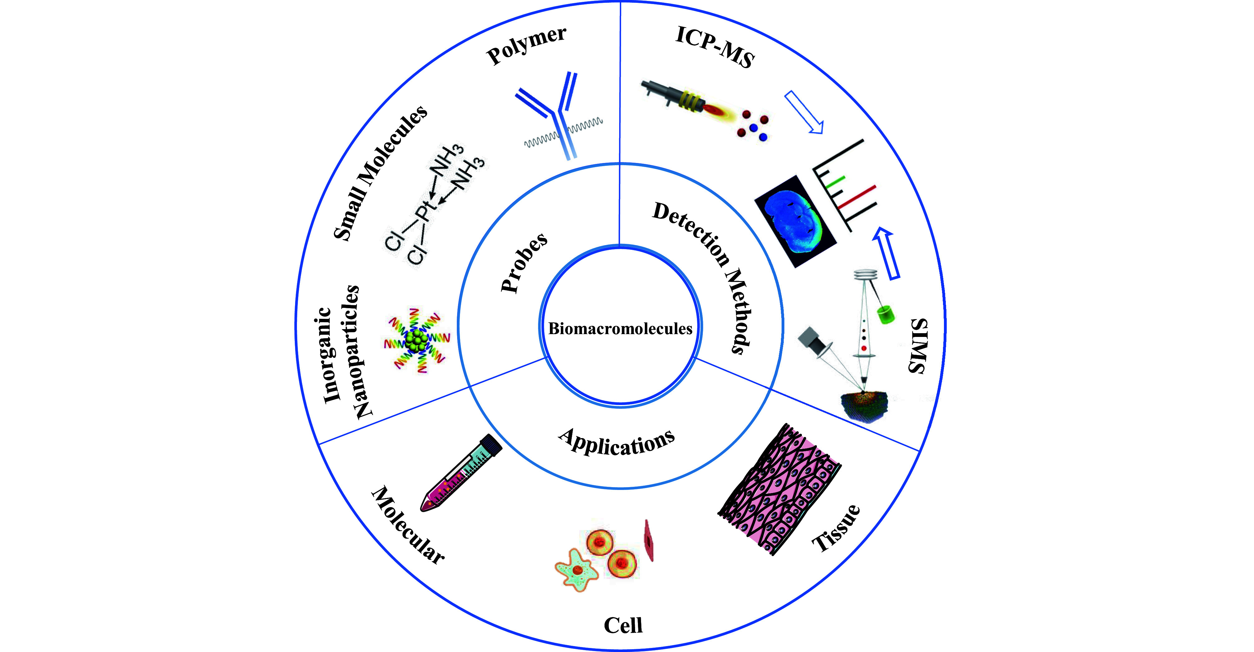
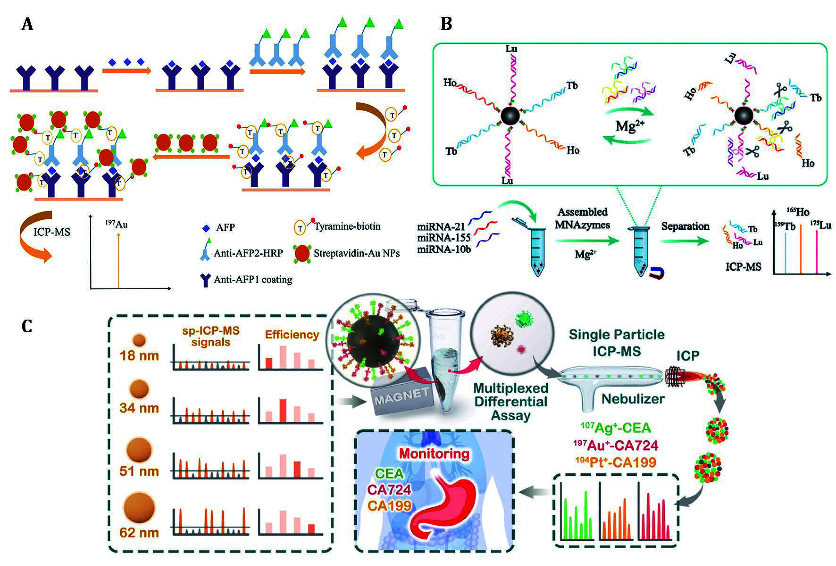
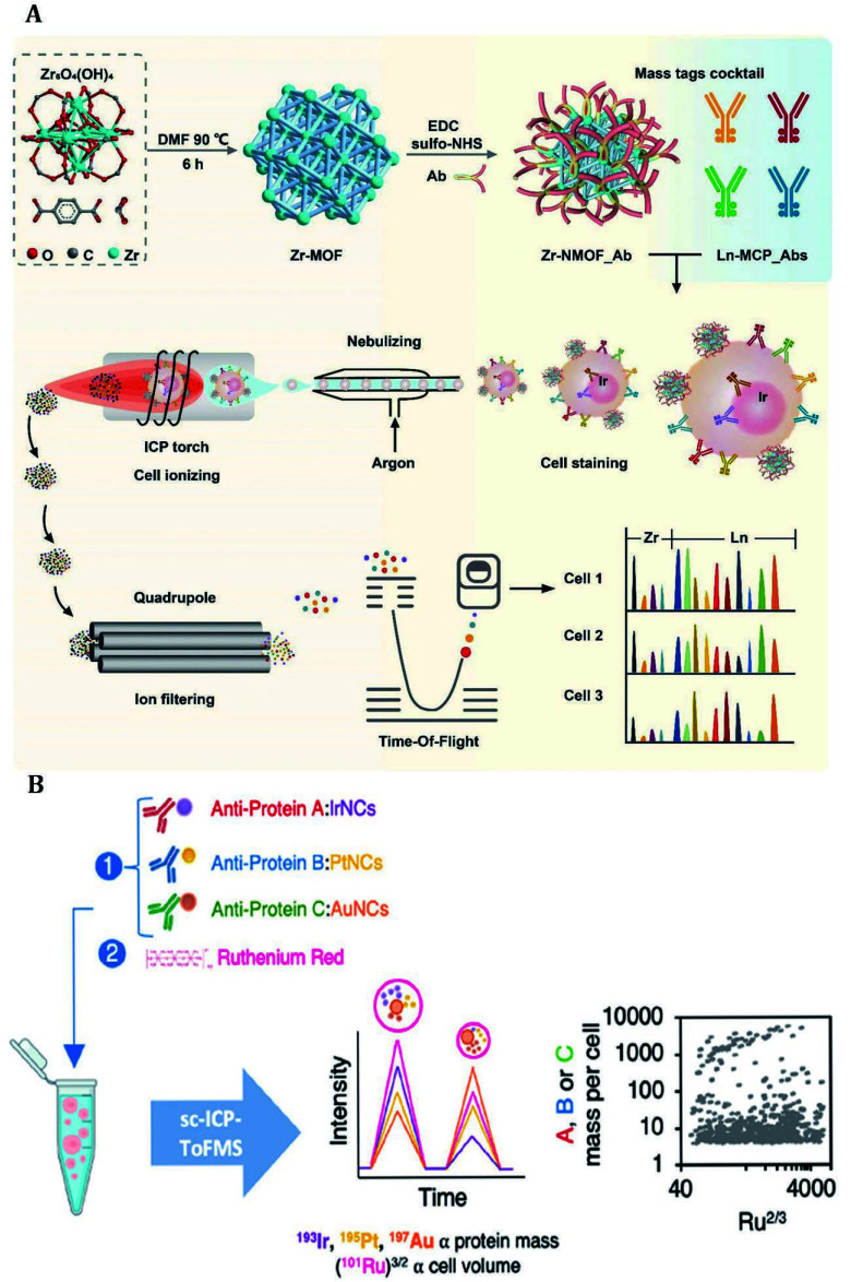
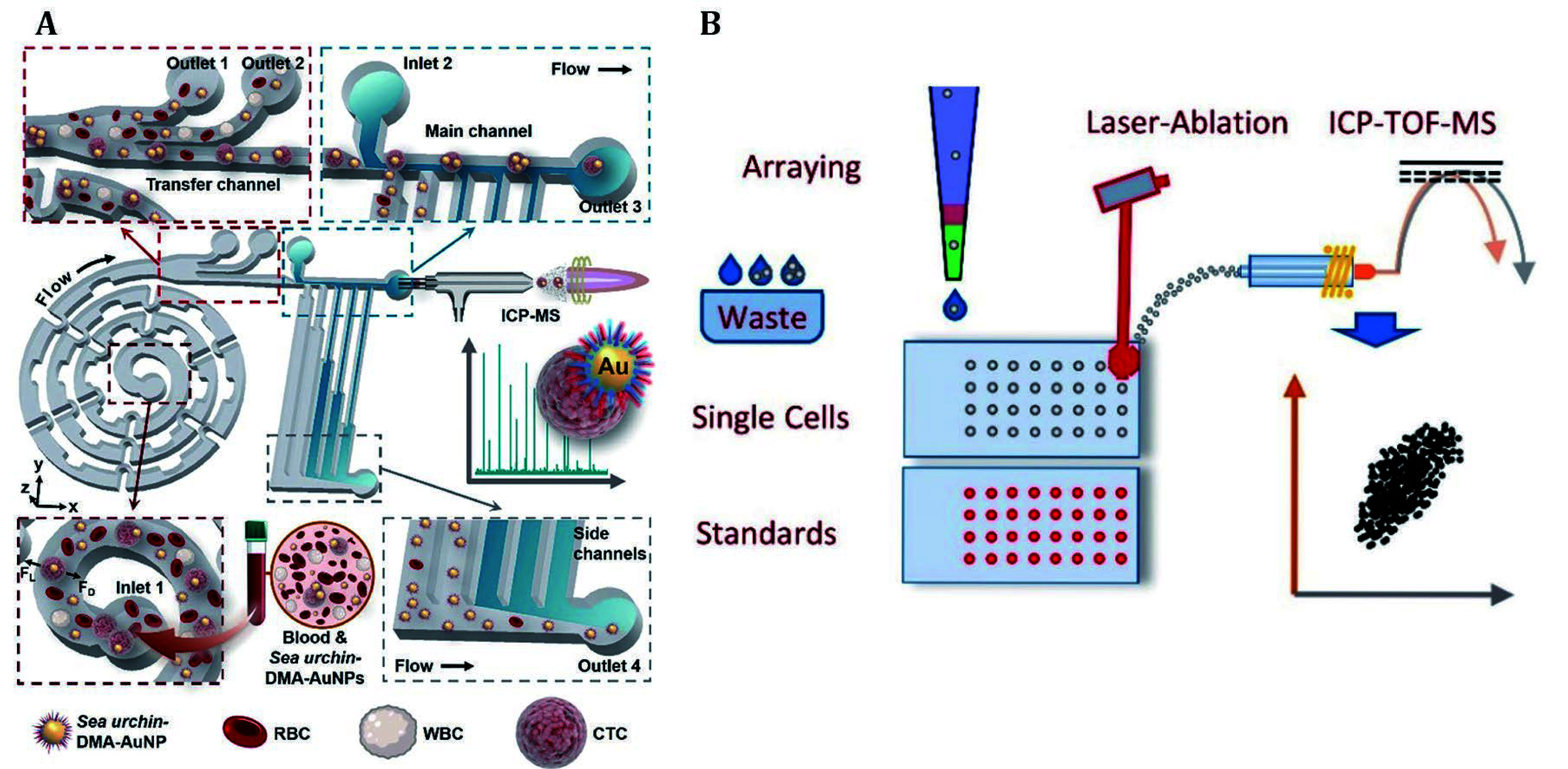
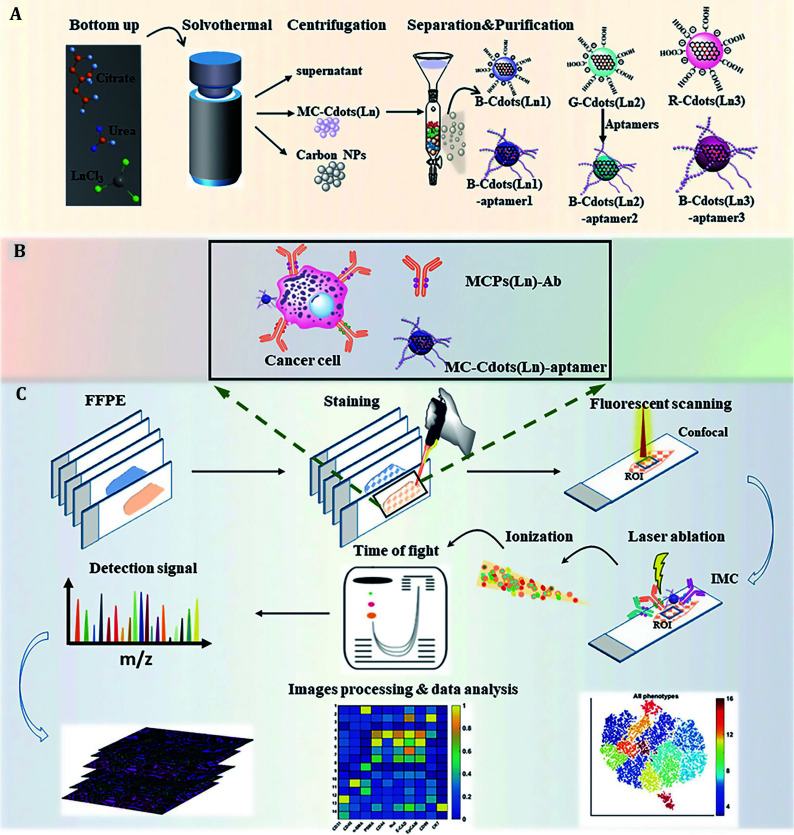
Biomacromolecules including proteins and nucleic acids are widely recognized for their pivotal and irreplaceable role in maintaining the normal functions of biological systems. By combining metal stable isotope labeling with elemental mass spectrometry, researchers can quantify the amount and track the spatial distribution of specific biomacromolecules in complex biological systems. In this review, the probes classification and metal stable isotope labeling strategies are initially summarized. Secondly, the technical characteristics and working principle of the elemental mass spectrometry techniques including inductively coupled plasma mass spectrometry and secondary ion mass spectrometry are introduced to achieve highly sensitive detection of multiple biomacromolecules at molecular, cellular and tissue levels. Lastly, we underline the advantages and limitations of elemental mass spectrometry combined with metal stable isotope labeling strategies, and propose the perspectives for future developments.

Keywords: Biomacromolecule; Mass spectrometry analysis; Metal stable isotope labeling.


Conflict of interest statement

Ping Zhang, Ying Han, Yue Xu and Liang Gao declare that they have no conflict of interest.

Figures

Figure 1
Figure 1
The applications of elemental mass spectrometry techniques combined with metal stable isotopes labeling for the quantification of biomolecules at molecular, cellular, and tissue levels are outlined
Figure 2
Figure 2
Enhanced detection sensitivity of free protein and miRNA through amplification strategies. A Free AFP was evaluated by the amplified elemental signal that was obtained by catalyzing the deposition of biotinylated tyramine and coupling with gold nanoparticles (Li et al. 2018). B The MNAzyme amplification strategy was combined with Ln elements labeling to enable sensitive detection of three miRNAs (Kang et al. 2021). C Three tumor biomarkers in the biological fluid were determined by evaluating the concentration of metal nanoparticles before and after immunoreaction with magnetic microspheres using SP-ICP-MS (Huang et al. 2022)
Figure 3
Figure 3
A Zr-NMOF was synthesized and bioconjugated with antibodies by EDC/sulfo-NHS reaction. Zr-NMOF targeting CD45, together with MCP-Abs targeting CD19, CD3, CD4, and CD8 were used for cell staining, while DNA labeling was accomplished through iridium insertion. High-temperature plasma evaporation of the samples generated element ion clouds for identification and qualitative analysis of cellular biomarkers by TOF-MS (Dang et al. 2021). B By labeling ferritin, metallothionein-2, and transferrin with antibodies decorated metal clusters, the relative concentration of proteins in individual cells was directly accessed by using RR staining to determine individual cell volume (Menero-Valdés et al. 2023)
Figure 4
Figure 4
A The sea urchin-inspired single-cell CTC recognition platform was developed, which captured CTCs by incubating sea urchin-DMA-AuNPs with blood. The CTCs were then separated from other cells and free probes in a chip based on secondary flow and hydrodynamic filtration. Subsequently, individual CTCs were subjected to background-free single-cell analysis by time-resolved ICP-MS (Zhang et al. 2021). B The pressure-acoustic single-cell microarray was developed for the detection of endogenous and exogenous isotopes within cells by LA-ICP-TOF-MS (Löhr et al. 2019)
Figure 5
Figure 5
The protocol began with nucleic acid amplification and peroxidase-catalyzed hapten deposition, followed by the labeling of nucleic acid and protein targets on sliced tissues. Subsequently, multiplexed images were acquired by following MIBI and CODEX platforms, and computational analysis was conducted to elucidate the host-pathogen interactions with high resolution (Jiang et al. 2022)
Figure 6
Figure 6
The strategy employed bimodal probes for the rapid identification and detection of ROI, thereby reducing the time of MS scanning. A The synthesis and purification of MC-Cdots (Ln) were performed to prepare bimodal probes. B A combined library consisting of the aptamer labeled MC-Cdots (Ln) and Maxpar antibody reagents was utilized for sample labeling. C MC-Cdots (Ln)-aptamer acted as a bimodal probe for the rapid identification of ROI through fluorescence signal, facilitating multiplex detection of ROI on the same tissue section by IMC (Yu et al. 2021)

Similar articles

References

    1. Agüi-Gonzalez P, Dankovich TM, Rizzoli SO, Phan NTN Gold-conjugated nanobodies for targeted imaging using high-resolution secondary ion mass spectrometry. Nanomaterials. 2021;11:1797. doi: 10.3390/nano11071797. - DOI - PMC - PubMed
    1. Allo B, Lou X, Bouzekri A, Ornatsky O Clickable and high-sensitivity metal-containing tags for mass cytometry. Bioconjugate Chem. 2018;29:2028–2038. doi: 10.1021/acs.bioconjchem.8b00239. - DOI - PubMed
    1. Angelo M, Bendall SC, Finck R, Hale MB, Hitzman C, Borowsky AD, Levenson RM, Lowe JB, Liu SD, Zhao S, Natkunam Y, Nolan GP Multiplexed ion beam imaging of human breast tumors. Nat Med. 2014;20:436–442. doi: 10.1038/nm.3488. - DOI - PMC - PubMed
    1. Anyaegbu CC, Lee-Pullen TF, Miller TJ, Abel TN, Platell CF, McCoy MJ Optimisation of multiplex immunofluorescence for a non-spectral fluorescence scanning system. J Immunol Methods. 2019;472:25–34. doi: 10.1016/j.jim.2019.06.011. - DOI - PubMed
    1. Arnett LP, Rana R, Chung WW, Li X, Abtahi M, Majonis D, Bassan J, Nitz M, Winnik MA Reagents for mass cytometry. Chem Rev. 2023;123:1166–1205. doi: 10.1021/acs.chemrev.2c00350. - DOI - PubMed

LinkOut - more resources