. 2025 May 1;135(9):e182709.
doi: 10.1172/JCI182709.

CXCL12+ fibroblastic reticular cells in lymph nodes facilitate immune tolerance by regulating T cell-mediated alloimmunity

Affiliations

CXCL12+ fibroblastic reticular cells in lymph nodes facilitate immune tolerance by regulating T cell-mediated alloimmunity

Yuta Yamamura et al. J Clin Invest. .

Abstract

Fibroblastic reticular cells (FRCs) are the master regulators of the lymph node (LN) microenvironment. However, the role of specific FRC subsets in controlling alloimmune responses remains to be studied. Single-cell RNA sequencing (scRNA-Seq) of naive and draining LNs (DLNs) of heart-transplanted mice and human LNs revealed a specific subset of CXCL12hi FRCs that expressed high levels of lymphotoxin-β receptor (LTβR) and are enriched in the expression of immunoregulatory genes. CXCL12hi FRCs had high expression of CCL19, CCL21, indoleamine 2,3-dioxygenase (IDO), IL-10, and TGF-β1. Adoptive transfer of ex vivo-expanded FRCs resulted in their homing to LNs and induced immunosuppressive environments in DLNs to promote heart allograft acceptance. Genetic deletion of LTβR and Cxcl12 in FRCs increased alloreactivity, abrogating the effect of costimulatory blockade in prolonging heart allograft survival. As compared with WT recipients, CXCL12+ FRC-deficient recipients exhibited increased differentiation of CD4+ T cells into Th1 cells. Nano delivery of CXCL12 to DLNs improved allograft survival in heart-transplanted mice. Our study highlights the importance of DLN CXCL12hi FRCs in promoting transplant tolerance.

Keywords: Adaptive immunity; Immunology; Tolerance; Transplantation.


Figures

Figure 1
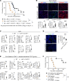
Figure 1. Adoptive transfer of FRCs promotes immune regulatory environment in DLNs.
(A) T cell activation assay of CD3+ T cells cocultured with or without WT-FRCs. Percentage of effector T cells evaluated by flow cytometry. (B) Th differentiation assay of CD4+ T cells cocultured with or without WT-FRC administration. Percentage of CD4+T-bet+ cells, and CD4+Rorγt+ cells evaluated by flow cytometry. (C) Chemotaxis assay of T cells with CM from cultured WT-FRCs. (D) Representative images of LNs injected with FRCs from Ccl19CretdTomato mice. Scale bars: 100 μm. (E) Intravital imaging of LNs injected with DsRed+ FRCs into Chst4-GFP mice. Yellow arrows indicate injected FRCs inside HEVs. (F) Representative images of LNs injected with FRC from DsRed mice. Yellow arrows indicate the FRCs contacting Foxp3+ cells. Scale bars: 100 μm. (G) Representative images and comparison of injected DsRed+ T cell homing in LNs with or without FRCs (n = 3–4 mice/group). (H) Representative images and comparison of Foxp3-stained DLNs from WT C57BL/6J recipients with or without FRCs after heart transplantation (n = 5 mice/group). Scale bars: 100 μm. (I) Flow cytometry analysis of DLNs from WT C57BL/6J recipients with or without FRC administration after heart transplantation (n = 5 mice/group). Student’s t test for comparisons between 2 groups. Data are represented as means ± SEM. *P < 0.05; **P < 0.01; ***P < 0.001; ****P < 0.0001.
Figure 2
Figure 2. Deletion of LTβR+ signaling on FRCs abrogates transplant tolerance.
(A) Comparison of heart allograft survival among WT C57BL/6J, Ccl19CreLtbrfl/fl, and Ccl19CreLtbrfl/fl recipients with FRC injections (n = 7–9 mice/group). Graft survival data were combined from 3 independent experiments. (B) Representative images and comparison of CD3-, CD11b-, and collagen 1–stained heart allografts from WT C57BL/6J and Ccl19CreLtbrfl/fl recipients (n = 4 mice/group). Scale bars: 200 μm. (C) Flow cytometry analysis of DLNs from WT C57BL/6J and Ccl19CreLtbrfl/fl recipients (n = 4 mice/group). (D) Representative images and comparison of Foxp3-stained DLNs (n = 4 mice/group). Scale bars: 200 μm. (E) Flow cytometry analysis of transferred TCR Tg cells in DLNs (n = 4 mice/group). (F) Comparison of heart allograft survival among WT C57BL/6J recipients treated with anti-LTβR agonist mAb alone (20 μg i.v. on day 0), anti-CD40L alone (40 μg i.v. on day 0), and both anti-LTβR agonist mAb and anti-CD40L of BALB/c hearts (n = 6 mice/group). Graft survival data were combined from 2 independent experiments. log-rank test for graft survival. Student’s t test for comparisons between 2 groups. Data are represented as means ± SEM. *P < 0.05; **P < 0.01; ***P < 0.001; ****P < 0.0001.
Figure 3
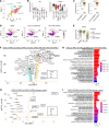
Figure 3. CXCL12hi FRC subsets enriched for genes for T cell migration.
(A) Unsupervised clustering of FRC subset clusters visualized with UMAP in LNSCs from naive LNs. (B) Violin plots of Ltbr and Cxcl12 expression among FRC subset clusters in naive LNs. (C) Violin plots of Ltbr and Cxcl12 expression in total FRCs from naive LNs. (D) UMAP of SC populations in naive LNs and DLNs, highlighting Cxcl12 expression on MedRC, TRC, and PRC subsets. (E) Violin plots of Cxcl12 expression in FRC subsets from naive LNs and DLNs. (F) Volcano plot comparing CXCL12hi and CXCL12lo FRCs in mouse LNSCs from heart transplanted recipients with anti-CD40L treatment. (G) Top 20 overrepresented ontology pathways based on DEGs in mouse LNSCs. (H) Volcano plot comparing CXCL12hi and CXCL12lo FRCs in human LNSCs. (I) Top 20 overrepresented ontology pathways in human LNSCs. Common genes shared between mouse and human are highlighted with bold letters surrounded by squares (F and H). Ontology pathways related to chemotaxis and migration are highlighted with bold letters surrounded by squares (G and I). HTx, heart transplantation.
Figure 4
Figure 4. Ablation of CXCL12 of FRCs abrogates anti-CD40L mediated transplant tolerance.
(A) Flow cytometry analysis of T cell compartments in naive LNs (n = 3 mice/group). (B) Representative intravital images of CMFDA-labeled CD3+ T cells injected into naive LNs. Yellow and white arrows indicate the migration of transferred CMFDA+ CD3+ T cells from inside HEVs into the parenchyma. Representative trajectories of transferred CD3+ T cells (gray, cyan, and magenta lines) and quantitative analysis of cell migration from inside HEVs toward the parenchyma for 20 minutes (n = 3). Scale bars: 100 μm. (C) In vivo T cell migration assay in a skin transplantation model. Representative images and quantitative analysis of CMFDA-labeled CD3+ T cells in DLNs (n = 5). Scale bars: 100 μm. (D) Comparison of heart allograft survival among the recipients under CTLA4 Ig treatment (n = 7 mice/group). Graft survival data were combined from 2 independent experiments. (E) Representative images and comparison of CD3-, CD11c-, and collagen 1–stained heart allografts (n = 5 mice/group). Scale bars: 200 μm. (F) Flow cytometry analysis of DLNs (n = 5 mice/group). log-rank test for graft survival. Student’s t test for comparisons between 2 groups. Data are represented as means ± SEM. *P < 0.05; **P < 0.01; ***P < 0.001; ****P < 0.0001.
Figure 5
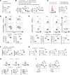
Figure 5. Deletion of CXCL12 in FRCs reduces Treg migration and immune suppressive effects for activated T cells.
(A) Comparison of CXCL12 expression in supernatants of FRC culture 24 hours after incubation (n = 3/group). (B) Chemotaxis assay of CD4+ T cells toward CM from WT-FRCs or CXCL12-KO-FRCs (n = 3/group). (C) Comparison of CXCR4 expression between CD4+CD25Foxp3 and CD4+CD25+Foxp3+ T cells (n = 3/group). (D) T cell activation assay of CD3+ T cells cocultured with WT-FRCs or CXCL12-KO-FRCs (n = 3/group). (E) Differentiation of CD4+ T cells into Th1, Th2, Th17, and Treg subsets in the presence of WT-FRCs or CXCL12-KO-FRCs (n = 3/group). (F) mRNA expression of IFN-γ and granzyme b in CD3+ T cells cocultured with WT-FRCs or CXCL12-KO-FRCs (n = 3/group). (G) Comparison of IDO, TGF-β1, IL-10, and PD-L1 expression between CXCL12lo and CXCL12hi FRCs in mouse LNs (n = 3/group). (H) Comparison of IDO, IL-10, and TGF-β1 expression between CXCL12lo and CXCL12hi FRCs in human LNs (n = 3/group). (I) Flow cytometry analysis of CD4+ Th1 differentiation with or without FRCs. CD4+ T cells were pretreated with DMSO, CXCR4 antagonist, or pertussis toxin for 1 hour (n = 3/group). Student’s t test for comparisons between 2 groups. One-way ANOVA with Tukey’s multiple-comparisons test for multiple comparisons. Data are represented as means ± SEM. *P < 0.05; **P < 0.01; ***P < 0.001; ****P < 0.0001.
Figure 6
Figure 6. Nano delivery of CXCL12 to LNs prolongs heart allograft survival.
(A) Schematic of Alexa Fluor 594–CXCL12–MECA79–NP synthesis. (B) Hydrodynamic size of MECA79-CXCL12-NPs in dynamic light-scattering analysis. (C) Loading efficiency of CXCL12 in NPs. (D) Release kinetics of CXCL12-NPs. (E) Ex vivo fluorescence imaging of DLNs from skin allograft recipients injected i.v. with free Alexa Fluor 594–CXCL12 or Alexa Fluor 594–CXCL12–MECA–79–NPs (n = 4 mice/group). (F) Representative images of DLNs 24 hours after i.v. injection of Alexa Fluor 594–CXCL12–MECA79–NPs. Scale bars: 100 μm (G) Comparison of heart allograft survival among recipient groups (n = 4–6 mice/group). (H) Representative images of heart allografts (n = 3 mice/group). Scale bars: 200 μm. log-rank test for graft survival. Student’s t test for comparisons between 2 groups. Data are represented as means ± SEM. *P < 0.05; **P < 0.01; ***P < 0.001.

References

    1. Ochando JC, et al. Lymph node occupancy is required for the peripheral development of alloantigen-specific Foxp3+ regulatory T cells. J Immunol. 2005;174(11):6993–7005. doi: 10.4049/jimmunol.174.11.6993. - DOI - PubMed
    1. Li L, et al. The lymph node stromal laminin α5 shapes alloimmunity. J Clin Invest. 2020;130(5):2602–2619. doi: 10.1172/JCI135099. - DOI - PMC - PubMed
    1. Li L, et al. Lymph node fibroblastic reticular cells preserve a tolerogenic niche in allograft transplantation through laminin α4. J Clin Invest. 2022;132(13):e156994. doi: 10.1172/JCI156994. - DOI - PMC - PubMed
    1. Li X, et al. Lymph node fibroblastic reticular cells deposit fibrosis-associated collagen following organ transplantation. J Clin Invest. 2020;130(8):4182–4194. - PMC - PubMed
    1. Ochando JC, et al. Direct versus indirect allorecognition: Visualization of dendritic cell distribution and interactions during rejection and tolerization. Am J Transplant. 2006;6(10):2488–2496. doi: 10.1111/j.1600-6143.2006.01494.x. - DOI - PubMed

LinkOut - more resources