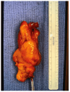
Congenital Malignant Ectomesenchymoma Presenting as a Neck Mass in a Newborn
- PMID: 40310147
- PMCID: PMC12025713
- DOI: 10.3390/children12040480
Congenital Malignant Ectomesenchymoma Presenting as a Neck Mass in a Newborn
Abstract
Background: Malignant Ectomesenchymoma (MEM) is a rare, aggressive soft tissue neoplasm with both neuroectodermal and mesenchymal differentiation. Congenital cases are extremely uncommon, posing significant diagnostic and therapeutic challenges. Case Presentation: We report a case of a full-term male neonate presenting with a large congenital neck mass and respiratory distress at birth. Imaging revealed a lobulated, heterogeneously enhancing mass in the left submandibular region with a mass effect on the airway. Open biopsy and gross resection on day six of life confirmed MEM with rhabdomyoblastic and neuroectodermal differentiation. Post-surgical staging classified the tumor as Stage I, Clinical Group II. Despite initial chemotherapy with Vincristine, Actinomycin, and Cyclophosphamide (VAC), tumor recurrence was detected at week nine of chemotherapy, necessitating a transition to Vincristine, Irinotecan, and Temozolomide (VIT). Discussion: MEM is an extremely rare neoplasm in infants, particularly in congenital presentations. Diagnosis is challenging due to its mixed histopathological features and broad differential diagnosis, including rhabdomyosarcoma, fibrosarcoma, lymphangioma, and neuroblastoma. Management typically involves multimodal therapy, with surgical resection being the mainstay of treatment. Chemotherapy is often tailored to the tumor's most aggressive component, though standardized treatment protocols remain undefined. Conclusions: This case highlights the importance of early recognition and a multidisciplinary approach in managing congenital MEM, as a differential diagnosis of soft tissue masses in infants, particularly in the head and neck region.
Keywords: congenital neck mass; malignant ectomesenchymoma; neonatology; newborn; soft tissue neoplasm.
Conflict of interest statement
The authors declare no conflicts of interest.
Figures


Similar articles
-
Malignant ectomesenchymoma in children and adolescents: report from the Cooperative Weichteilsarkom Studiengruppe (CWS).Pediatr Blood Cancer. 2013 Feb;60(2):224-9. doi: 10.1002/pbc.24174. Epub 2012 Apr 25. Pediatr Blood Cancer. 2013. PMID: 22535600
-
Disseminated malignant ectomesenchymoma (MEM): case report and review of the literature.Pediatr Hematol Oncol. 2002 Jan-Feb;19(1):9-17. doi: 10.1080/088800102753356149. Pediatr Hematol Oncol. 2002. PMID: 11787870 Review.
-
More Than Meets the Eye? A Cautionary Tale of Malignant Ectomesenchymoma Treated as Low-risk Orbital Rhabdomyosarcoma.J Pediatr Hematol Oncol. 2021 Aug 1;43(6):e854-e858. doi: 10.1097/MPH.0000000000001901. J Pediatr Hematol Oncol. 2021. PMID: 32769567
-
An integrative morpho-molecular approach in malignant ectomesenchymoma diagnosis: report of a new paediatric case and a review of the literature.Front Oncol. 2024 Mar 1;14:1320541. doi: 10.3389/fonc.2024.1320541. eCollection 2024. Front Oncol. 2024. PMID: 38496756 Free PMC article. Review.
-
A Complex Case of Diffused Malignant Extra-Renal Rhabdoid Tumor in a Newborn.Ear Nose Throat J. 2025 Mar 19:1455613251325041. doi: 10.1177/01455613251325041. Online ahead of print. Ear Nose Throat J. 2025. PMID: 40107999
References
-
- Pellegrino F., Tirtei E., Divincenzo F., Campello A., Rubino C., Augustoni E., Linari A., Asaftei S.D., Fagioli F. An integrative morpho-molecular approach in malignant ectomesenchymoma diagnosis: Report of a new paediatric case and a review of the literature. Front. Oncol. 2024;14:1320541. doi: 10.3389/fonc.2024.1320541. - DOI - PMC - PubMed
-
- Milano G.M., Orbach D., Casanova M., Berlanga P., Schoot R.A., Corradini N., Brennan B., Ramirez-Villar G.L., Hjalgrim L.L., van Noesel M.M., et al. Malignant ectomesenchymoma in children: The European pediatric Soft tissue sarcoma Study Group experience. Pediatr. Blood Cancer. 2023;70:e30116. - PubMed
Publication types
LinkOut - more resources
Full Text Sources

