

Optimal Wavelengths for Multispectral Short Wavelength Infrared Transillumination and Reflectance Imaging for Caries Detection
- PMID: 40310441
- PMCID: PMC12026095
- DOI: 10.3390/diagnostics15081034
Optimal Wavelengths for Multispectral Short Wavelength Infrared Transillumination and Reflectance Imaging for Caries Detection
Abstract
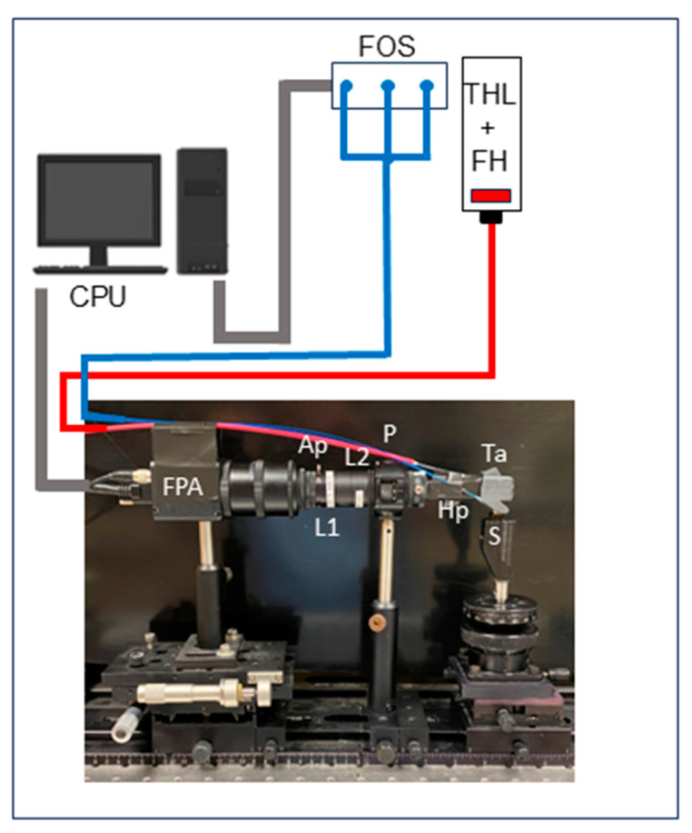
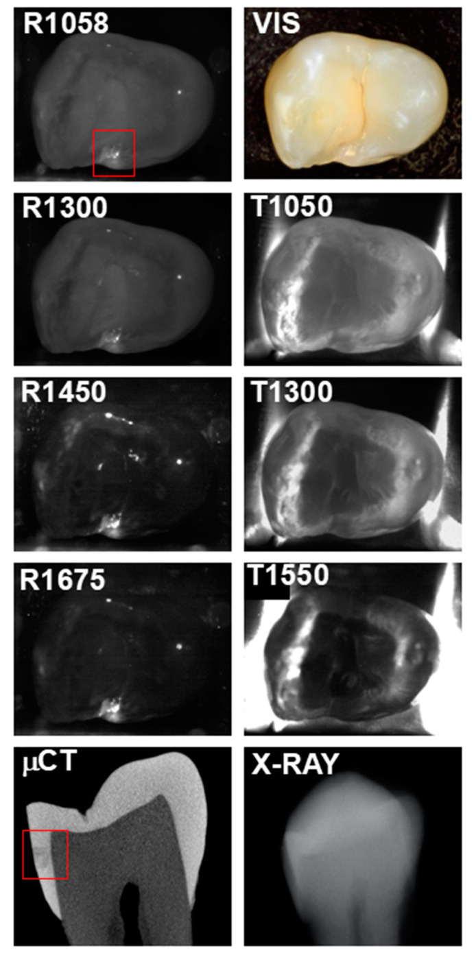
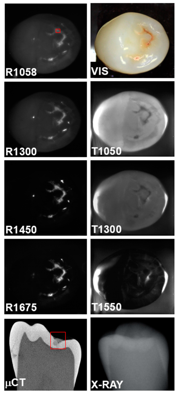
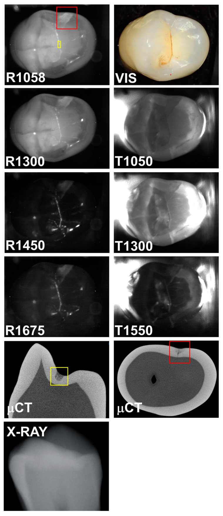
Background/Objectives: The aim of this in vitro study was to determine the optimal combinations of wavelengths for short wavelength infrared (SWIR) multispectral transillumination and reflectance imaging of caries lesions on proximal and occlusal surfaces. Methods: The contrasts of (n = 76) caries lesions on the occlusal and proximal surfaces of extracted teeth were measured at 1050, 1300, and 1550 nm for occlusal transillumination and 1058, 1300, 1450, and 1675 nm for occlusal reflectance. All teeth were also imaged using radiography and microcomputed tomography (μCT) to verify lesion presence. A custom-fabricated handheld imaging probe suitable for clinical use and for the simultaneous acquisition of SWIR occlusal transillumination and reflectance (SWIR-OTR) images was used. Three high-power superluminescent diode lasers were used for transillumination, and a fiber-optic switch was used to switch between the transillumination wavelengths. Optical bandpass filters coupled with a tungsten halogen lamp were used for reflectance. All images were acquired at the same position and with the same field of view for comparison. Results: The highest contrasts in reflection were at 1450 and 1675 nm for occlusal and interproximal lesions, and the highest contrasts for transillumination were at 1050 and 1300 nm. Conclusions: This study suggests that the best wavelengths for SWIR-OTR are between 1000 and 1300 nm for transillumination and greater than 1400 nm for reflectance. Wavelengths beyond 1400 nm are advantageous for reflectance and yield significantly higher contrast. Wavelengths beyond 1300 nm are not promising for occlusal transillumination since internal water absorption leads to contrast inversion.
Keywords: SWIR imaging; dental caries; reflectance imaging; transillumination.
Conflict of interest statement
The authors declare no conflicts of interest.
Figures
References
-
- NIDCR . Oral Health in America: A Report of the Surgeon General-Executive Summary. U.S. Department of Health and Human Services, National Institute of Dental and Craniofacial Research, National Institutes of Health; Rockville, MD, USA: 2020.
-
- Makhija S.K., Gilbert G.H., Funkhouser E., Bader J.D., Gordan V.V., Rindal D.B., Bauer M., Pihlstrom D.J., Qvist V., National Dental Practice-Based Research Network Collaborative Group The prevalence of questionable occlusal caries: Findings from the Dental Practice-Based Research Network. J. Am. Dent. Assoc. 2012;143:1343–1350. doi: 10.14219/jada.archive.2012.0097. - DOI - PMC - PubMed
-
- Fried D. SWIR Imaging of lesions on Tooth Surfaces. In: Sordillo L.A., Sordillo D.C., editors. Short-Wavelength Infrared Windows for Biomedical Applications. SPIE; Bellingham, WA, USA: 2022. pp. 521–543.
Grants and funding
LinkOut - more resources
Full Text Sources
