. 2025 May 6;122(18):e2423842122.
doi: 10.1073/pnas.2423842122. Epub 2025 May 1.

Next-generation rifamycins for the treatment of mycobacterial infections

Affiliations

Next-generation rifamycins for the treatment of mycobacterial infections

Véronique Dartois et al. Proc Natl Acad Sci U S A. .

Abstract

Mycobacterium abscessus is a rapidly growing nontuberculous Mycobacterium causing severe pulmonary infections, especially in immunocompromised individuals and patients with underlying lung conditions like cystic fibrosis (CF). While rifamycins are the pillar of tuberculosis treatment, their efficacy against M. abscessus lung disease is severely compromised by intrabacterial ADP-ribosylation. Additionally, rifamycins induce cytochrome P450 3A4 (CYP3A4), a major human drug-metabolizing enzyme, further limiting their use in patients with comorbidities that require treatment with CYP3A4 substrates such as CF and HIV coinfection. We chemically reengineered rifabutin to enhance its potency against M. abscessus by blocking intrabacterial inactivation and eliminate drug-drug interactions by removing induction of CYP3A4 gene expression. We have designed and profiled a series of C25-substituted derivatives resistant to intracellular inactivation and lacking CYP3A4 induction, while retaining excellent pharmacological properties. Against Mycobacterium tuberculosis, devoid of ADP-ribosyltransferase, the frontrunners are equipotent to rifabutin, suggesting superior clinical utility since they no longer come with the drug interaction liability typical of rifamycins. Prioritized compounds demonstrated superior antibacterial activity against a panel of M. abscessus clinical isolates, were highly bactericidal against replicating and drug-tolerant nonreplicating bacteria in caseum surrogate and were active against intracellular bacteria. As single agents, these rifamycins were as effective as a standard-of-care four-drug combination in a murine model of M. abscessus lung infection.

Keywords: Mycobacterium abscessus; drug discovery; preclinical development candidate; pulmonary infection; rifamycin.


Conflict of interest statement

Competing interests statement:PCT/US2023/034573 (16).

Figures

Fig. 1.
Fig. 1.
Rifamycin screening cascade and optimized leads. (A) Left: rifabutin scaffold showing the C23 position where ADP-ribosylation occurs and the C25 position where substituents are introduced to block Arr-mediated ribosylation. Right: screening and compound profiling cascade including threshold metrics. PPB: plasma protein binding, fu: fraction unbound; AUC: area under the concentration–time curve; PDCs: preclinical development candidates. (B) Potency and C25 substituents of the six selected leads that meet pharmacokinetic–pharmacodynamic criteria. MICs (concentrations that inhibit 90% growth) against the wild-type Mab reference strain ATCC19977 (MICWT) and isogenic Δarr knockout strain (MICΔarr). UMN-22 and UMN-46 are referred to as 5m and 5n in ref. , respectively
Fig. 2.
Fig. 2.
Impact of cLogP on in vitro PK parameters and cytotoxicity. (A) cLogP versus unbound fraction (fu) in mouse and human plasma for the series of RBT analogs. (B) Impact of cLogP on uptake into THP-1 macrophages and concentration that induces 50% HepG2 cell death (CC50).
Fig. 3.
Fig. 3.
Bactericidal activity of the frontrunners against nonreplicating Mab in caseum surrogate (A) and intracellular Mab ATCC19977 in THP-1-derived macrophages (B). (A) The CFU/mL caseum are expressed in percent of starting burden, with MBC90 indicating 90% kill over a 5-d treatment duration. Clarithromycin (CLR) was used as control and RBT as the rifamycin comparator. (B) Drug treatment was initiated 2 h post infection for 48 h, at 1 and 10 µM. Assays were carried out in technical triplicates. In all 1- and 10-µM treatment groups with RBT analogs, intracellular bacterial burden was significantly lower than prior to treatment initiation (2 h), indicating bactericidal activity (one-way ANOVA with Dunnett’s posttest, all P values < 0.0001 **** not shown for clarity). Dose–response was queried by comparing the 1- and 10-µM treatment groups using Student’s t test. ns: not significant, **: P < 0.01. UT: untreated; DMSO: vehicle only.
Fig. 4.
Fig. 4.
Efficacy of the selected rifamycin leads in the GM-CSF−/− mouse model of acute Mab infection and PK-PD profiles of the preclinical development candidates. (A) Reduction of lung CFU in GM-CSF−/− mice following seven oral daily doses administered between Day 2 and Day 9 (D2, D9) at 25 mg/kg except UMN-123 and UMN-132 given at 12.5 mg/kg. LOD: limit of detection or 1.6 log CFU. (B) Efficacy of a typical intensive phase drug combination (amikacin AMK, imipenem IMP, clofazimine CFZ, and tedizolid TED) administered for 7 d at human-equivalent doses. (C) Dose–response efficacy of the selected PDCs UMN-120 and UMN-121 from 2 to 25 mg/kg as indicated. D9 DF: Drug-free arm on D9 after seven daily doses. Numbers at the bottom of each column are mean Log CFU/mouse (n = 5). (D) Plasma concentration–time profiles of RBT and the two preclinical development candidates in CD-1 mice following administration as indicated, compared to key potency values; dotted red line: MIC against Mab ATCC19977, solid red line: serum-shifted MIC; green line: MIC90 or minimum concentration that inhibits growth of 90% of clinical isolates (n = 76).
Fig. 5.
Fig. 5.
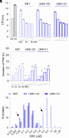
Extensive microbiological profiling of the PDCs. (A) Dose–response bactericidal activity in replicating cultures at multiples of the MIC against the type strain ATCC19977, measured after 3 d (D3) of incubation. (B) Post-antibiotic effect (PAE) of RBT and the PDCs at multiples of their respective MICs. (C) MIC distributions of RBT and the PDCs against a panel of 76 M. abscessus clinical isolates. The arrows indicate the position of the M. abscessus subsp. abscessus type strain ATCC19977.

References

    1. Prideaux B., et al. , The association between sterilizing activity and drug distribution into tuberculosis lesions. Nat. Med. 21, 1223–1227 (2015). - PMC - PubMed
    1. Xie M., et al. , ADP-ribosylation-resistant rifabutin analogs show improved bactericidal activity against drug-tolerant M. abscessus in caseum surrogate. Antimicrob. Agents. Chemother. 67, e0038123 (2023), 10.1128/aac.00381-23. - DOI - PMC - PubMed
    1. Fox W., Ellard G. A., Mitchison D. A., Studies on the treatment of tuberculosis undertaken by the British Medical Research Council tuberculosis units, 1946–1986, with relevant subsequent publications. Int. J. Tuberc. Lung Dis. 3, S231–279 (1999). - PubMed
    1. Loeliger A., et al. , Protease inhibitor-containing antiretroviral treatment and tuberculosis: Can rifabutin fill the breach? Int. J. Tuberc Lung Dis. 16, 6–15 (2012). - PubMed
    1. Dorman S. E., et al. , Four-month rifapentine regimens with or without moxifloxacin for tuberculosis. N. Engl. J. Med. 384, 1705–1718 (2021). - PMC - PubMed

MeSH terms

LinkOut - more resources