

Assessment of metal artifacts from titanium wrist prostheses: photon-counting versus energy-integrating detector CT
- PMID: 40310571
- PMCID: PMC12045920
- DOI: 10.1186/s41747-025-00587-w
Assessment of metal artifacts from titanium wrist prostheses: photon-counting versus energy-integrating detector CT
Abstract
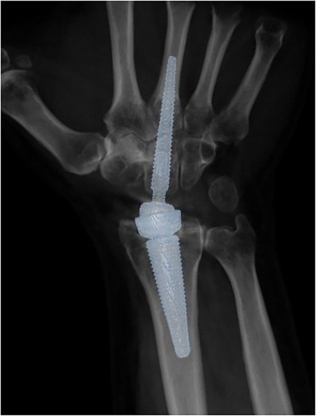
Background: We compared photon-counting detector computed tomography (PCD-CT) polyenergetic images, PCD-CT virtual monoenergetic images (VMI), and energy-integrating detector computed tomography (EID-CT) polyenergetic images regarding bone visualization and metal artifacts in patients with titanium wrist prostheses.
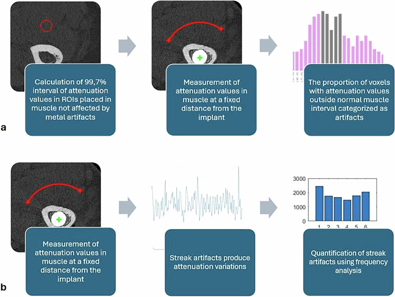
Methods: After ethical approval, 15 patients were examined with PCD-CT and EID-CT. Polyenergetic images were reconstructed, as well as 130-keV VMI for PCD-CT. Five radiologists evaluated bone visualization, interpretability at metal-bone interface and metal artifacts using a 7-point ordinal scale. Streak artifacts and artifacts at the bone-metal interface were quantitatively assessed. Differences between image setups were analyzed using Friedman test and one-way ANOVA with post hoc tests.
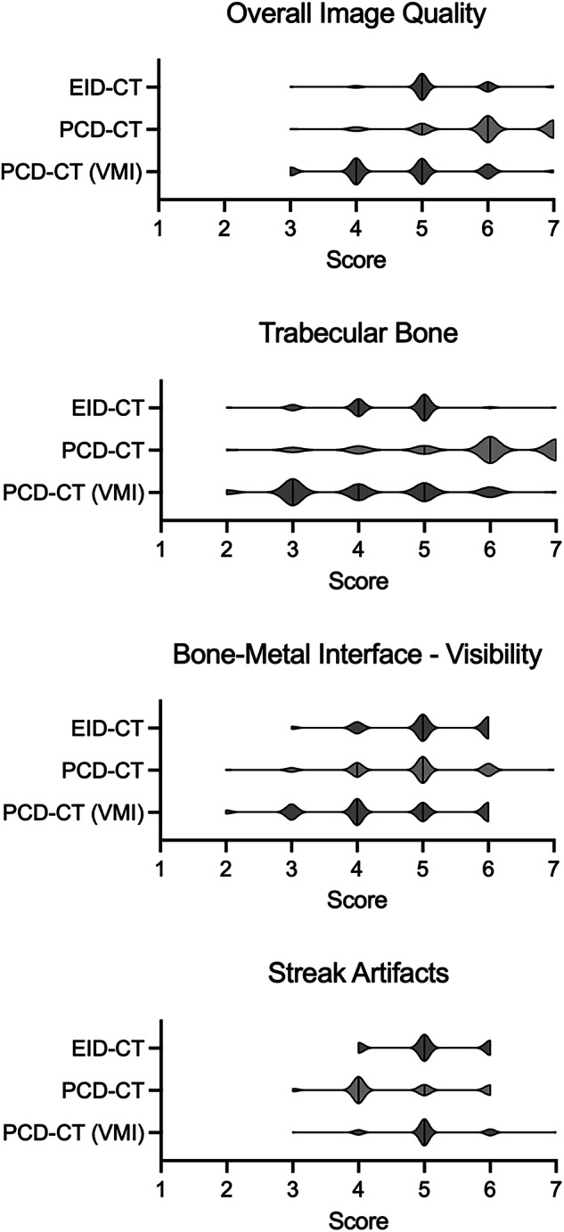
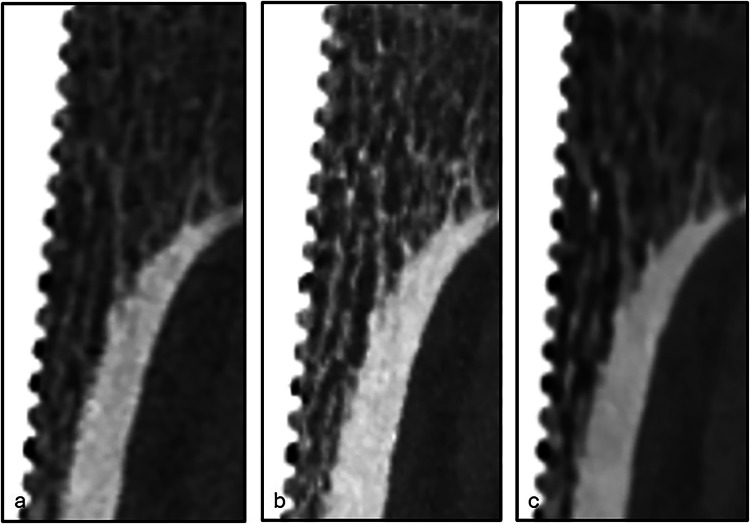
Results: Bone visualization was superior in PCD-CT polyenergetic images (median rating 6, range 3-7) compared with VMI (5, 3-7; p < 0.001) and EID-CT (5, 3-7; p = 0.018). Streak artifacts were more pronounced with PCD-CT polyenergetic images (4, 3-6) compared with EID-CT (5, 4-6; p = 0.003) and PCD-CT VMI (5, 3-7; p = 0.002), with quantitative results showing least streak artifacts in PCD-CT VMI, followed by EID-CT and PCD-CT polyenergetic images (50 ± 7%, 70 ± 6%, and 79 ± 5%, respectively; p < 0.001). Interpretability at bone-metal interface was better with PCD-CT polyenergetic images (5, 2-7; p = 0.045) and EID-CT (5, 3-6; p = 0.018) compared with PCD-CT VMI (4, 2-6), without quantitative differences.
Conclusion: Streak artifacts from titanium wrist prostheses were reduced using 130-keV PCD-CT VMI, while bone visualization was highest using PCD-CT polyenergetic images.
Relevance statement: In patients with wrist implants, photon-counting detector CT allows for effective metal artifact reduction using virtual monoenergetic images and improved bone visualization using polyenergetic images. As polyenergetic images and VMI have different advantages, access to both image setups may benefit diagnostic evaluation.
Key points: Virtual monoenergetic images (VMI) presented a substantial reduction of metal streak artifacts. Polyenergetic images exhibited better image quality for bone imaging compared with VMI. A combination of image reconstructions should be preferred depending on the diagnostic task.
Keywords: Arthroplasty; Artifacts; Titanium; Tomography (x-ray computed); Wrist joint.
© 2025. The Author(s).
Conflict of interest statement
Declarations. Ethics approval and consent to participate: The study was approved by the Swedish Ethical Review Authority (Dnr. 2021-02034). Consent for publication: Patients eligible to participate received oral and written information about the study and were given the option to decline. Competing interests: SF provides consulting services for Swemac Innovation AB. RB has been, since February 2025, employed as Scientific Product Manager at Siemens Healthineers. The remaining authors declare no conflicts of interest.
Figures
Similar articles
-
Combining virtual monoenergetic imaging and iterative metal artifact reduction in first-generation photon-counting computed tomography of patients with dental implants.Eur Radiol. 2023 Nov;33(11):7818-7829. doi: 10.1007/s00330-023-09790-y. Epub 2023 Jun 7. Eur Radiol. 2023. PMID: 37284870 Free PMC article.
-
Photon-Counting Detector CT: Clinical Utility of Virtual Monoenergetic Imaging Combined With Tin Prefiltration to Reduce Metal Artifacts in the Postoperative Ankle.Invest Radiol. 2024 Aug 1;59(8):545-553. doi: 10.1097/RLI.0000000000001058. Epub 2024 Jan 10. Invest Radiol. 2024. PMID: 38214560
-
Influence of virtual monoenergetic reconstructions on coronary CT angiography-based fractional flow reserve with photon-counting detector CT: intra-individual comparison with energy-integrating detector CT.Insights Imaging. 2025 Feb 17;16(1):36. doi: 10.1186/s13244-025-01927-5. Insights Imaging. 2025. PMID: 39961979 Free PMC article.
-
Photon-Counting Detector CT for Musculoskeletal Imaging: A Clinical Perspective.AJR Am J Roentgenol. 2023 Apr;220(4):551-560. doi: 10.2214/AJR.22.28418. Epub 2022 Oct 19. AJR Am J Roentgenol. 2023. PMID: 36259593 Review.
-
An introduction to photon-counting detector CT (PCD CT) for radiologists.Jpn J Radiol. 2023 Mar;41(3):266-282. doi: 10.1007/s11604-022-01350-6. Epub 2022 Oct 18. Jpn J Radiol. 2023. PMID: 36255601 Free PMC article. Review.
References
-
- Redfern JAI, Mehta N, Farnebo S et al (2024) Complication rates and modes of short and medium-term failure in Motec total wrist arthroplasty: an international cohort study. J Hand Surg Eur Vol 49:27–33. 10.1177/17531934231195689 - PubMed
-
- Boeckstyns MEH, Herzberg G (2024) Complications after total wrist arthroplasty. J Hand Surg Eur Vol 49:177–187. 10.1177/17531934231203297 - PubMed
-
- Barrett JF, Keat N (2004) Artifacts in CT: recognition and avoidance. Radiographics 24:1679–1691. 10.1148/rg.246045065 - PubMed
-
- Grandmougin A, Bakour O, Villani N et al (2020) Metal artifact reduction for small metal implants on CT: which image reconstruction algorithm performs better? Eur J Radiol 127:108970. 10.1016/j.ejrad.2020.108970 - PubMed
-
- Katsura M, Sato J, Akahane M, Kunimatsu A, Abe O (2018) Current and novel techniques for metal artifact reduction at CT: practical guide for radiologists. Radiographics 38:450–461. 10.1148/rg.2018170102 - PubMed
Publication types
MeSH terms
Substances
Grants and funding
LinkOut - more resources
Full Text Sources
Medical
Research Materials